Ch. 10 Muscle Tissue and Physiology
Amerman - Human Anatomy & Physiology 2nd Edition
Amerman2nd EditionHuman Anatomy & PhysiologyISBN: 9780136873822Not the one you use?Change textbook
Chapter 10, Problem 2

How does a skeletal muscle fiber differ structurally from typical cells?

Verified step by step guidance
1
Skeletal muscle fibers are much larger than typical cells, often extending the entire length of the muscle. This is due to their formation through the fusion of multiple embryonic cells called myoblasts, making them multinucleated.
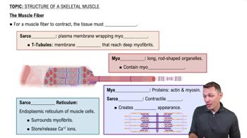
Unlike typical cells, skeletal muscle fibers are multinucleated, meaning they contain multiple nuclei. These nuclei are located at the periphery of the cell, just beneath the sarcolemma (the plasma membrane of the muscle fiber).
Skeletal muscle fibers have a specialized plasma membrane called the sarcolemma, which is involved in conducting electrical signals (action potentials) necessary for muscle contraction.
Inside skeletal muscle fibers, there are unique organelles called myofibrils, which are composed of repeating units called sarcomeres. Sarcomeres are the functional units of muscle contraction and are not found in typical cells.
Skeletal muscle fibers also contain a specialized endoplasmic reticulum called the sarcoplasmic reticulum, which stores and releases calcium ions (Ca²⁺) critical for muscle contraction. This is a structural adaptation not present in most typical cells.

Verified video answer for a similar problem:

This video solution was recommended by our tutors as helpful for the problem above.
Was this helpful?

Key Concepts

Here are the essential concepts you must grasp in order to answer the question correctly.

Skeletal Muscle Fiber Structure

Skeletal muscle fibers are unique in their structure, characterized by being long, cylindrical, and multinucleated. Unlike typical cells, which usually have a single nucleus, skeletal muscle fibers contain multiple nuclei located at the periphery of the cell. This structural adaptation allows for efficient coordination of muscle contraction and repair.
Recommended video:
Guided course
7:47
The Muscle Fiber

Striations in Muscle Fibers

Skeletal muscle fibers exhibit striations, which are alternating light and dark bands visible under a microscope. These striations result from the organized arrangement of myofibrils, which contain actin and myosin filaments. This highly organized structure is essential for the muscle's ability to contract efficiently and generate force.
Recommended video:
Guided course
7:47
The Muscle Fiber

Sarcomeres

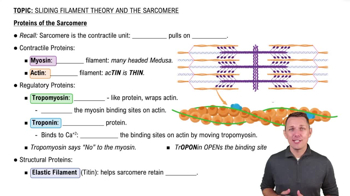
Sarcomeres are the fundamental contractile units of skeletal muscle fibers, defined by the region between two Z discs. Each sarcomere contains overlapping thick (myosin) and thin (actin) filaments, which slide past each other during muscle contraction. This sliding filament mechanism is crucial for muscle function and distinguishes skeletal muscle from other cell types.
Recommended video:
Guided course
7:18
Proteins of the Sarcomere
Related Practice
Textbook Question

Which type of muscle fascicle pattern has an appearance similar to a feather?

a. Fusiform

b. Triangular

c. Pennate

d. Parallel

851
views

Textbook Question

Mark the following statements as true or false. If a statement is false, correct it to make a true statement.

The plasma membrane of a muscle cell is called the sarcoplasmic reticulum.

823
views
Textbook Question

Mark the following statements as true or false. If a statement is false, correct it to make a true statement.

Muscle cells are contractile, conductive, distensible cells.

811
views
Textbook Question

The primary action of the biceps brachii muscle of the anterior arm is to flex the forearm at the elbow. However, when this muscle is inflamed, pain is felt in the shoulder. Explain this finding.

880
views
Textbook Question

Jesse is a 2-year-old boy who presents with difficulty in walking and poor control of movements. When the doctor examines Jesse, she notices that when his muscles contract, they are very slow to relax and remain contracted well after the movement has been performed. She sends a sample of his tissue for genetic analysis, and the lab reports a genetic defect that causes the pumps in the SR to operate much more slowly than normal. How does a defect in DNA lead to a malfunctioning protein? How does this finding explain Jesse's symptoms?

753
views
Textbook Question

Ms. Sanchez was in a motorcycle accident in which she lost the use of her right upper limb muscles due to significant nerve damage. However, when an electrode is inserted into her muscles, they are able to contract. Explain specifically why nerve damage caused her to lose the use of her muscles. Why can they still respond to stimulation from an electrode?

781
views