Immersion oil ________(increases/decreases) the numerical aperture, which ________(increases/decreases) resolution because _______(more/fewer) light rays are involved.
Ch. 4 - Microscopy, Staining, and Classification
Chapter 4, Problem 4.4a
________ refers to differences in intensity between two objects.
 Verified step by step guidance
1
Identify the context in which the term is used, as it could relate to various fields such as microscopy, imaging, or general scientific observation.
In the context of microscopy, consider how differences in intensity can affect the visibility and differentiation of structures within a sample.
Understand that in microscopy, the term often refers to 'contrast,' which is crucial for distinguishing between different parts of a specimen.
Recognize that contrast can be enhanced through various techniques, such as staining, phase contrast, or differential interference contrast.
Consider how improving contrast can lead to better visualization and interpretation of microscopic images, aiding in the identification and study of microorganisms.

Verified Solution
Video duration:
1mWas this helpful?
Key Concepts
Here are the essential concepts you must grasp in order to answer the question correctly.
Contrast
Contrast in microbiology refers to the differences in intensity or color between two objects, such as cells or tissues, when viewed under a microscope. It is crucial for distinguishing between different structures and components in a sample, allowing for better visualization and analysis of microbial organisms.
Recommended video:
Guided course

Magnification, Resolution, & Contrast
Microscopy Techniques
Various microscopy techniques, such as bright-field, dark-field, and phase-contrast microscopy, utilize contrast to enhance the visibility of specimens. Each technique manipulates light differently to improve the contrast between the specimen and its background, aiding in the observation of microbial morphology and behavior.
Recommended video:
Guided course

Light Microscopy: Bright-Field Microscopes
Staining Methods
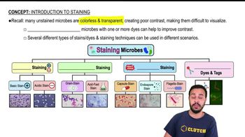
Staining methods are used to increase contrast in microbiological samples by adding dyes that bind to specific cellular components. Techniques like Gram staining or acid-fast staining help differentiate between types of bacteria based on their cell wall properties, enhancing the contrast and making it easier to identify and classify microorganisms.
Recommended video:
Guided course

Introduction to Staining
Related Practice
Textbook Question
80
views
Textbook Question
Why can electron microscopes magnify only dead organisms?
84
views
Textbook Question
Curved glass lenses _______light.
a. refract
b. bend
c. magnify
d. both a and b
77
views
Textbook Question
Put the following substances in the order they are used in a Gram stain: counterstain, decolorizing agent, mordant, primary stain.
56
views
Textbook Question
Which of the following factors is important in making an image appear larger?
a. thickness of the lens
b. curvature of the lens
c. speed of the light passing through the lens
d. all of the above
79
views
Textbook Question
Cationic chromophores such as methylene blue ionically bond to _______(positively/negatively) charged chemicals such as DNA and proteins.
85
views
