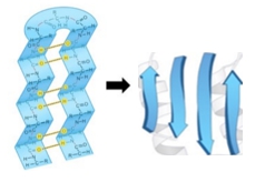
So now that we've talked about alpha helices, we can move on and talk about our next type of secondary structure, the beta strand. The beta strand, again, is a type of secondary structure, and this is a structure where the protein backbone takes an extended zigzag conformation that is periodic and repeats. By zigzag, what you'll see is, if you take a quick look at our example down below, that the R groups are zigzagging. When you look at the R groups which are in green, they go from being down to going up to going down to going up to going down, and so what you'll see is that the R groups are literally zigzagging, and that's what we mean by the zigzag conformation. The extended periodic zigzag conformation or structure repeats every 2 amino acid residues. The rise, so if we take a look at the rise and remember that the rise is just the length or the distance covered per amino acid residue, is actually about 3.5 Ångströms, and if we compare this to the rise of an alpha helix, that's actually more than double because the rise of an alpha helix is just 1.5 Ångströms. So, basically, what that's saying is that the beta strand is much more extended. It's more extended than the alpha helix, which is much more coiled together. The pitch of a beta strand is going to be 7 Ångströms, and again, that's much longer than the pitch of an alpha helix which is just 5.4 Ångströms. All that's saying is that the beta strand is more extended than the alpha helix which is coiled. In our example below, we're going to compare the rise, pitch, and length of 5 amino acid residues in a beta strand conformation and then 5 amino acid residues in an alpha helix conformation. In our example down below, we have the beta strand on the left over here and the alpha helix on the right. On the far left over here, what we have is our key: the green represent the R groups, the red balls represent oxygens, blue are the nitrogens, black are carbons, and white are hydrogens. Comparing the beta strand to the alpha helix, they both have exactly 5 amino acid residues. One of the first things that you'll notice is that the beta strand conformation on the left over here is much more extended. The backbone for our beta strand is literally just much more extended, whereas with the alpha helix, it's much more coiled together. The rise for a beta strand is 3.5 Ångströms, whereas the rise for an alpha helix is just 1.5 Ångströms. Notice that the distance here, the distance of the arrow, this arrow here and this arrow over here, is just much, much more extended. One single amino acid residue stretches out much further and extends more in the beta strand conformation. Now, looking at the pitch, for an alpha helix it is when you have one turn of the alpha helix backbone. Here, the pitch is referring to this blue line here and this blue line over here, and that's exactly where there's one turn of the backbone. The pitch comes out to 5.4 Ångströms in the alpha helix. Now, for the pitch in a beta strand, it's referring to these lines over here, showing the repeated structure of the beta strand. Notice that the R groups are both going down here, and the distance between this periodic repeat structure is just 2 amino acid residues, but the length turns out to be 7 Ångströms, and that's what the pitch actually is. The way that we calculate the length of a beta strand is going to be the same way that we calculated the length for the alpha helix. All we needed to do was take the number of amino acid residues and multiply it by the rise. For this beta strand here, which has 5 amino acid residues, all we need to do is take 5 and multiply it by the rise of 3.5 Ångströms. That comes out to 17.5 Ångströms, and that's the length of this beta strand over here on the left. If we do the same for the alpha helix, which again has 5 amino acid residues, we'll take the 5 amino acid residues and multiply it by 1.5 Ångströms because that's the rise for the alpha helix. This ends up coming out to just 7.5 Ångströms. What you can see is that when you're comparing 5 amino acids in a beta strand, notice that the length is 17.5 Ångströms, which is much longer and extended than 5 amino acids in an alpha helix conformation, which is coiled. That's really the biggest thing that you want to take away from this, along with the pitch and the rise and the ability to calculate the lengths here. And so, that concludes our lesson on beta strands, and I'll see you guys in our practice.
Beta Strand - Online Tutor, Practice Problems & Exam Prep
Beta Strand
Video transcript
What is the approximate length of a β-strand containing 27 amino acids?
Beta Strand
Video transcript
So now that we know that the beta strand is just an extended zigzag conformation of the protein backbone, we can talk about beta strand depictions. Beta strands are commonly depicted as extended broad arrows, these arrows can actually twist, and they point towards the C-terminal end of the protein. Similar to alpha helices, the beta strands stabilize the protein structure. However, unlike alpha helices, the hydrogen bonds are not parallel; they are perpendicular to the direction of the beta strands. Recall that perpendicular means that when they intersect, they form 90-degree angles.
Looking at our example, we will label the terminals of the beta strand. On the left, we have a single beta strand, represented by one arrow. This arrow points towards the C-terminal end, indicating that this must be the C-terminal end, and consequently, the opposite end must be the N-terminal end. This is straightforward because we always consider sequences from the N-terminal end to the C-terminal end. Having the arrow point towards the C-terminal end seems quite natural and should be easy for you to remember.
On the right, we have two beta strands, each indicated by an arrow. These two arrows are connected into a single chain, both pointing towards their respective C-terminal ends. Thus, the end where the tip of the arrow is located must be the C-terminal end of this protein, and the end where the back of the arrow is located has to be the N-terminal end. When beta strands are aligned this way, they are stabilized by hydrogen bonds of the peptide backbone, which we will discuss in more detail later in our course. For now, what you need to know is that these hydrogen bonds are perpendicular to the directions of these strands, aligning almost perpendicular to the direction of the strands, which is a significant difference from alpha helices.
This concludes our lesson on beta strand depictions, and we will get a little bit of practice in our next video, so I'll see you there.
Which phrase best describes the hydrogen bonds of a β-strand in silk fibroin, a protein with β-conformations?