Ch. 3 - Observing Microorganisms Through a Microscope
Chapter 3, Problem 3.6a

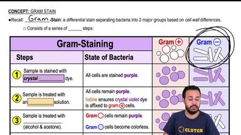
Why is a mordant used in the Gram stain? In the flagella stain?

Verified step by step guidance
1
Understand that a mordant is a substance used to set or stabilize stains or dyes.
In the Gram stain, the mordant (iodine) is used to form a complex with the crystal violet dye, which helps to fix the dye within the cell wall of Gram-positive bacteria.
Recognize that the iodine-crystal violet complex is larger and more insoluble than the crystal violet alone, making it less likely to be washed out during the decolorization step.
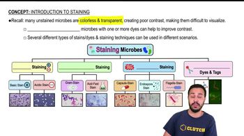
In the flagella stain, the mordant is used to increase the diameter of the flagella, making them visible under a light microscope by binding to the stain and the flagella.
The mordant in the flagella stain helps to coat the flagella with the stain, allowing for better visualization of these thin structures.

Verified Solution

Video duration:
1m
This video solution was recommended by our tutors as helpful for the problem above.
Was this helpful?

Key Concepts

Here are the essential concepts you must grasp in order to answer the question correctly.

Mordant in Gram Staining

A mordant is a chemical agent used in the Gram staining process to enhance the binding of the primary stain, crystal violet, to the bacterial cell wall. It typically forms a complex with the dye, making it less soluble and more firmly attached to the peptidoglycan layer. This is crucial for differentiating between Gram-positive and Gram-negative bacteria, as the former retains the dye after decolorization.
Recommended video:

Flagella Staining

In flagella staining, a mordant is used to increase the thickness of the flagella, making them more visible under a microscope. Since flagella are thin and difficult to see, the mordant helps to precipitate the dye onto the flagella, allowing for better visualization of their structure and arrangement. This is essential for identifying motility in bacteria.
Recommended video:
Guided course
02:10
Introduction to Staining

Differential Staining Techniques

Differential staining techniques, such as Gram staining and flagella staining, are used to distinguish between different types of bacteria based on their structural characteristics. These methods rely on the use of specific dyes and mordants to highlight differences in cell wall composition or appendages. Understanding these techniques is fundamental in microbiology for classifying and identifying bacterial species.
Recommended video:
Guided course
03:27
Differential Staining
Related Practice
Textbook Question

Looking at the cell of a photosynthetic microorganism, you observe the chloroplasts are green in brightfield microscopy and red in fluorescence microscopy. You conclude:

a. chlorophyll is fluorescent.

b. the magnification has distorted the image.

c. you’re not looking at the same structure in both microscopes.

d. the stain masked the green color.

e. none of the above

118
views
Textbook Question

The maximum magnification of a compound microscope is (a) ________; that of an electron microscope, (b) ________. The maximum resolution of a compound microscope is (c) ________; that of an electron microscope, (d) ________. One advantage of a scanning electron microscope over a transmission electron microscope is (e) ________.

130
views
Textbook Question

Which of the following is not a functionally analogous pair of stains?

a. nigrosin and malachite green

b. crystal violet and carbolfuchsin

c. safranin and methylene blue

d. ethanol-acetone and acid-alcohol

e. All of the above pairs are functionally analogous.

118
views
Textbook Question

Which of the following pairs is mismatched?

a. capsule—negative stain

b. cell arrangement—simple stain

c. cell size—negative stain

d. Gram stain—bacterial identification

e. none of the above

189
views
Textbook Question

What is the purpose of a counterstain in the acid-fast stain?

134
views
Textbook Question

Assume you stain Clostridium by applying a basic stain, carbolfuchsin, with heat, decolorizing with acid-alcohol, and counterstaining with an acidic stain, nigrosin. Through the microscope, the endospores are 1, and the cells are stained 2.

a. 1—red; 2—black

b. 1—black; 2—colorless

c. 1—colorless; 2—black

d. 1—red; 2—colorless

e. 1—black; 2—red

141
views