Ch. 1 - Introduction to Microbiology
Norman-McKay- Microbiology: Basic and Clinical Principles 2nd Edition
Norman-McKay2nd EditionMicrobiology: Basic and Clinical PrinciplesISBN: 9780137661619Not the one you use?Change textbook
Chapter 1, Problem 14

Classify the following as bright field, dark field, phase contrast, or differential interference contrast (DIC).
a. Sample must be stained or have its own coloration to be seen:
b. Specimen is illuminated with polarized light:
c. Generates a falsely three-dimensional image:
d. The simplest and most common form of light microscopy:
e. Generates a negative image using a hollow cone of light:
f. Generates a negative image in which light shifts are converted into visible changes in brightness and contrast:

Verified step by step guidance
1
Step 1: Understand the characteristics of each microscopy technique: Bright field microscopy requires stained or naturally pigmented samples to provide contrast; dark field microscopy uses a hollow cone of light to create a bright image on a dark background, producing a negative image; phase contrast microscopy converts phase shifts in light passing through a transparent specimen into brightness changes; differential interference contrast (DIC) microscopy uses polarized light and Nomarski prisms to generate images with a three-dimensional appearance.
Step 2: For part (a), identify the technique that requires the sample to be stained or naturally colored to be visible. This is a key feature of bright field microscopy.
Step 3: For part (b), determine which microscopy method uses polarized light to illuminate the specimen. This is characteristic of differential interference contrast (DIC) microscopy.
Step 4: For part (c), find the technique that produces a falsely three-dimensional image, which is a hallmark of DIC microscopy due to its use of polarized light and interference.
Step 5: For parts (d) and (e), recognize that the simplest and most common form of light microscopy is bright field, while the method that generates a negative image using a hollow cone of light is dark field microscopy. For part (f), identify the technique that converts light phase shifts into visible brightness and contrast changes, which is phase contrast microscopy.

Verified video answer for a similar problem:

This video solution was recommended by our tutors as helpful for the problem above.
Video duration:
2m
Was this helpful?

Key Concepts

Here are the essential concepts you must grasp in order to answer the question correctly.

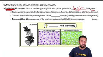
Bright Field Microscopy

Bright field microscopy is the most common light microscopy technique where the specimen is illuminated from below and observed against a bright background. Samples often require staining or must have natural coloration to be visible, as contrast depends on light absorption by the specimen.
Recommended video:
Guided course
03:46
Light Microscopy: Bright-Field Microscopes

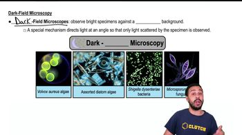
Dark Field Microscopy

Dark field microscopy uses a hollow cone of light to illuminate the specimen, which scatters light into the objective lens, producing a bright image on a dark background. This technique generates a negative image and is useful for observing live, unstained samples with enhanced contrast.
Recommended video:
Guided course
01:52
Dark-Field Microscopy

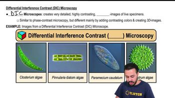
Phase Contrast and Differential Interference Contrast (DIC) Microscopy

Phase contrast microscopy converts phase shifts in light passing through transparent specimens into brightness changes, enhancing contrast without staining. DIC microscopy uses polarized light and optical gradients to produce high-contrast, pseudo three-dimensional images, revealing fine structural details in live cells.
Recommended video:
Guided course
02:03
Differential Interference Contrast (DIC) Microscopy