From memory, draw two eukaryotic cells. Label the structures listed here and show any physical connections between the internal structures of each cell: nucleus, rough ER, smooth ER, mitochondrion, centrosome, chloroplast, vacuole, lysosome, microtubule, cell wall, ECM, microfilament, Golgi apparatus, intermediate filament, plasma membrane, peroxisome, ribosome, nucleolus, nuclear pore, vesicle, flagellum, microvilli, plasmodesma.
Verified step by step guidance
1
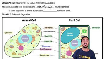
Start by drawing the outline of two eukaryotic cells. One should represent a typical animal cell, and the other a plant cell. This will help in distinguishing structures unique to each type, such as chloroplasts and cell walls in plant cells.
Inside each cell, draw the nucleus as a large, central organelle. Label the nucleolus inside the nucleus and indicate nuclear pores on the nuclear envelope. These pores are crucial for the transport of molecules between the nucleus and cytoplasm.
Draw the endoplasmic reticulum (ER) near the nucleus. The rough ER should have ribosomes on its surface, while the smooth ER should be depicted without ribosomes. These structures are involved in protein and lipid synthesis, respectively.
Illustrate the Golgi apparatus as a series of stacked membranes near the ER. This organelle is responsible for modifying, sorting, and packaging proteins and lipids for secretion or use within the cell.
Add other organelles and structures: mitochondria (energy production), centrosomes (microtubule organization), chloroplasts (photosynthesis, in plant cells), vacuoles (storage, larger in plant cells), lysosomes (digestion), microtubules, microfilaments, intermediate filaments (cytoskeleton components), plasma membrane (cell boundary), peroxisomes (metabolism), ribosomes (protein synthesis), vesicles (transport), flagellum (movement, in some animal cells), microvilli (surface area increase, in some animal cells), plasmodesma (communication between plant cells), cell wall (structure, in plant cells), and ECM (extracellular matrix, in animal cells). Label each structure clearly.
Verified video answer for a similar problem:
This video solution was recommended by our tutors as helpful for the problem above.
Video duration:
3m
Play a video:
Was this helpful?
Key Concepts
Here are the essential concepts you must grasp in order to answer the question correctly.
Eukaryotic Cell Structure
Eukaryotic cells are characterized by their complex structure, including a defined nucleus and various organelles. Key components include the nucleus, which houses genetic material, and organelles like mitochondria, ER, and Golgi apparatus, each performing specific functions. Understanding the layout and function of these structures is crucial for drawing and labeling eukaryotic cells accurately.
Each organelle within a eukaryotic cell has distinct roles, such as energy production by mitochondria or protein synthesis by ribosomes. These organelles often interact physically and functionally, such as the rough ER's role in protein synthesis and transport to the Golgi apparatus. Recognizing these interactions helps in illustrating connections between cell structures.
Beyond organelles, eukaryotic cells contain various structural components like microtubules, microfilaments, and intermediate filaments, which provide support and facilitate movement. The plasma membrane and cell wall define the cell's boundary, while structures like the ECM and plasmodesma are involved in cell communication and adhesion. Understanding these components is essential for comprehensive cell diagrams.
 Verified step by step guidance
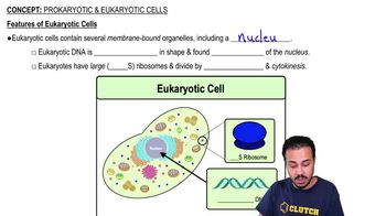
 04:17
04:17