Every skeletal muscle basically has two ends where it attaches to the skeleton, and when the muscle contracts, those two ends are going to get closer to each other. We're going to define those two ends as the origin and the insertion. So you can kind of think of the job of a muscle is to make two parts of the body get closer to each other. And when we do that, we're going to get the origin and the insertion closer. And specifically, we can say that the insertion, we're going to say the origin is the stationary attachment site. It doesn't move. The insertion, that's the mobile attachment site. That's the attachment site that moves. To see what we're talking about here, we have a diagram showing the bicep brachii. Now, bicep brachii, that's that sort of major muscle of your upper arm and has two attachment points. One's up in the shoulder and one's down here on the forearm. Well, if I flex my bicep brachii, my forearm is going to come up, my elbow is going to bend, and my forearm is going to keep on getting closer to my shoulder. The insertion moved closer to the origin. That means that the insertion must be down here on my forearm, and the origin didn't move, that's up in my shoulder. A little memory tool to keep these straight in your head, you can say the origin observes and the insertion inches closer. Now, learning the origin and insertion can sometimes just feel like another thing on the list of facts you got to memorize, but if you understand how a muscle works, they kind of just make sense. If you know the origin and the insertion, you should be able to figure out the movement of the muscle. If you know the movement of the muscle, you can often figure out the origin and the insertion. That's because the origin and the insertion are going to determine the movement. And to see what I mean, we have some examples here. We're going to show flexion at the knee. So here, this person is bending their knee, and their lower leg is sort of bending up, and they're using their bicep femoris. This is a muscle of a hamstring. You can see this has some attachment points up here on this end. One attachment point's on the ischium, and the other's on the back of the femur there. And then on the other end, it has an attachment point on the tibia and the fibula on the other side of the knee there. So, one way you can think of this movement of flexion is, well, you're getting your tibia and your fibula closer to your rear end. So, which end is moving? Well, down here on the tibia and fibula, that's moving. That's my insertion. I'm going to mark it with an I. The part that isn't moving, that's the origin. I'm going to mark it with an O. Next, we can look at extension. Extension is straightening the leg here. Sort of, straightening the leg at the knee. And we can see here we have a muscle that does that. That's the vastus intermedius. We have two attachment points for this muscle. One is on the front of the femur, and the other one sort of comes over the kneecap there, and attaches to the tibia at the tibial tuberosity. So, one way to think of extension is you're sort of getting the front of your tibia closer to the front of your femur. So, which part is moving? Well, that tibial tuberosity is moving, so that's going to be my insertion. The front of the femur is not moving, so that's going to be my origin. And I'll mark those. We can look at abduction. Abduction is taking an arm or a leg away from the midline of the body. So here we have the muscle, the gluteus medius, and this is attached up on the ilium and it's also attached on the greater trochanter. So, to pull the leg away like this, we can see here the greater trochanter of the femur. You can think of it as getting closer to the ilium. That's the job of this muscle. So, which part's moving? Well, the insertion moves. That's gotta be on the greater trochanter. I'll mark it with an I. The ilium does not move, so that's gotta be my origin. I'll mark it with an O. For adduction, adduction is bringing an arm or leg closer down into the midline of the body. Here, we're looking at the adductor longus muscle, and this attaches to the pubis and also to the femur down here. So, to bring this leg in towards the body, one way you can think of that, you're kinda taking the inside of your femur and moving it closer to the pubis. Well, which part's moving? The femur is moving, so that is going to be my insertion. The pubis isn't moving, so that's going to be my origin. Alright. Our last example here is going to be rotation. And for rotation, we're taking this leg and we're turning it out. So, you're sort of taking your foot and turning it out. And the muscle we're looking at here is the piriformis. And the piriformis has one attachment point on the sacrum and another on that greater trochanter, on the outside of the femur. So, one way you can think of this movement, that outside of the femur, that greater trochanter, is getting closer to the sacrum when you turn your leg out. That means that the insertion must be on that greater trochanter, and the origin must be on the sacrum. Alright. Again, trying to learn your origins and insertions of muscles on their own can sometimes just feel like a really unrelated list of facts. Remember, they cause the movements. So, if you know the origin and insertions, you know the movement. If you know the movement, you can usually figure out the origin and insertion.
- 1. Introduction to Anatomy & Physiology5h 40m
- What is Anatomy & Physiology?20m
- Levels of Organization13m
- Variation in Anatomy & Physiology12m
- Introduction to Organ Systems27m
- Homeostasis9m
- Feedback Loops11m
- Feedback Loops: Negative Feedback19m
- Feedback Loops: Positive Feedback11m
- Anatomical Position7m
- Introduction to Directional Terms3m
- Directional Terms: Up and Down9m
- Directional Terms: Front and Back6m
- Directional Terms: Body Sides12m
- Directional Terms: Limbs6m
- Directional Terms: Depth Within the Body4m
- Introduction to Anatomical Terms for Body Regions3m
- Anatomical Terms for the Head and Neck8m
- Anatomical Terms for the Front of the Trunk8m
- Anatomical Terms for the Back9m
- Anatomical Terms for the Arm and Hand9m
- Anatomical Terms for the Leg and Foot15m
- Review- Using Anatomical Terms and Directions12m
- Abdominopelvic Quadrants and Regions19m
- Anatomical Planes & Sections17m
- Organization of the Body: Body Cavities13m
- Organization of the Body: Serous Membranes14m
- Organization of the Body: Serous Membrane Locations8m
- Organization of the Body: Thoracic Cavity8m
- Organization of the Body: Abdominopelvic Cavity12m
- 2. Cell Chemistry & Cell Components12h 37m
- Atoms- Smallest Unit of Matter57m
- Isotopes39m
- Introduction to Chemical Bonding19m
- Covalent Bonds40m
- Noncovalent Bonds5m
- Ionic Bonding37m
- Hydrogen Bonding19m
- Introduction to Water7m
- Properties of Water- Cohesion and Adhesion7m
- Properties of Water- Density8m
- Properties of Water- Thermal14m
- Properties of Water- The Universal Solvent17m
- Acids and Bases12m
- pH Scale21m
- Carbon8m
- Functional Groups9m
- Introduction to Biomolecules2m
- Monomers & Polymers11m
- Carbohydrates23m
- Proteins25m
- Nucleic Acids34m
- Lipids28m
- Microscopes10m
- Prokaryotic & Eukaryotic Cells26m
- Introduction to Eukaryotic Organelles16m
- Endomembrane System: Protein Secretion34m
- Endomembrane System: Digestive Organelles15m
- Mitochondria & Chloroplasts21m
- Endosymbiotic Theory10m
- Introduction to the Cytoskeleton10m
- Cell Junctions8m
- Biological Membranes10m
- Types of Membrane Proteins7m
- Concentration Gradients and Diffusion9m
- Introduction to Membrane Transport14m
- Passive vs. Active Transport13m
- Osmosis33m
- Simple and Facilitated Diffusion17m
- Active Transport30m
- Endocytosis and Exocytosis15m
- 3. Energy & Cell Processes10h 7m
- Introduction to Energy15m
- Laws of Thermodynamics15m
- Chemical Reactions9m
- ATP20m
- Enzymes14m
- Enzyme Activation Energy9m
- Enzyme Binding Factors9m
- Enzyme Inhibition10m
- Introduction to Metabolism8m
- Redox Reactions15m
- Introduction to Cellular Respiration22m
- Types of Phosphorylation11m
- Glycolysis19m
- Pyruvate Oxidation8m
- Krebs Cycle16m
- Electron Transport Chain14m
- Chemiosmosis7m
- Review of Aerobic Cellular Respiration19m
- Fermentation & Anaerobic Respiration23m
- Introduction to Cell Division22m
- Organization of DNA in the Cell17m
- Introduction to the Cell Cycle7m
- Interphase18m
- Phases of Mitosis48m
- Cytokinesis16m
- Cell Cycle Regulation18m
- Review of the Cell Cycle7m
- Cancer13m
- Introduction to DNA Replication22m
- DNA Repair7m
- Central Dogma7m
- Introduction to Transcription20m
- Steps of Transcription19m
- Genetic Code25m
- Introduction to Translation30m
- Steps of Translation23m
- Post-Translational Modification6m
- 4. Tissues & Histology10h 3m
- Introduction to Tissues & Histology16m
- Introduction to Epithelial Tissue24m
- Characteristics of Epithelial Tissue37m
- Structural Naming of Epithelial Tissue19m
- Simple Epithelial Tissues1h 2m
- Stratified Epithelial Tissues55m
- Identifying Types of Epithelial Tissue32m
- Glandular Epithelial Tissue26m
- Introduction to Connective Tissue36m
- Classes of Connective Tissue8m
- Introduction to Connective Tissue Proper40m
- Connective Tissue Proper: Loose Connective Tissue56m
- Connective Tissue Proper: Dense Connective Tissue49m
- Specialized Connective Tissue: Cartilage44m
- Specialized Connective Tissue: Bone12m
- Specialized Connective Tissue: Blood9m
- Introduction to Muscle Tissue7m
- Types of Muscle Tissue45m
- Introduction to Nervous Tissue8m
- Nervous Tissue: The Neuron8m
- 5. Integumentary System2h 20m
- 6. Bones & Skeletal Tissue2h 16m
- An Introduction to Bone and Skeletal Tissue18m
- Gross Anatomy of Bone: Compact and Spongy Bone7m
- Gross Anatomy of Bone: Periosteum and Endosteum11m
- Gross Anatomy of Bone: Bone Marrow8m
- Gross Anatomy of Bone: Short, Flat, and Irregular Bones5m
- Gross Anatomy of Bones - Structure of a Long Bone23m
- Microscopic Anatomy of Bones - Bone Matrix9m
- Microscopic Anatomy of Bones - Bone Cells25m
- Microscopic Anatomy of Bones - The Osteon17m
- Microscopic Anatomy of Bones - Trabeculae9m
- 7. The Skeletal System2h 35m
- 8. Joints2h 17m
- 9. Muscle Tissue2h 33m
- 10. Muscles1h 11m
- 11. Nervous Tissue and Nervous System1h 35m
- 12. The Central Nervous System1h 6m
- 13. The Peripheral Nervous System1h 26m
- Introduction to the Peripheral Nervous System5m
- Organization of Sensory Pathways16m
- Introduction to Sensory Receptors5m
- Sensory Receptor Classification by Modality6m
- Sensory Receptor Classification by Location8m
- Proprioceptors7m
- Adaptation of Sensory Receptors8m
- Introduction to Reflex Arcs13m
- Reflex Arcs15m
- 14. The Autonomic Nervous System1h 38m
- 15. The Special Senses2h 41m
- 16. The Endocrine System2h 48m
- 17. The Blood1h 22m
- 18. The Heart1h 42m
- 19. The Blood Vessels3h 35m
- 20. The Lymphatic System3h 16m
- 21. The Immune System14h 37m
- Introduction to the Immune System10m
- Introduction to Innate Immunity17m
- Introduction to First-Line Defenses5m
- Physical Barriers in First-Line Defenses: Skin13m
- Physical Barriers in First-Line Defenses: Mucous Membrane9m
- First-Line Defenses: Chemical Barriers24m
- First-Line Defenses: Normal Microbiota7m
- Introduction to Cells of the Immune System15m
- Cells of the Immune System: Granulocytes28m
- Cells of the Immune System: Agranulocytes26m
- Introduction to Cell Communication5m
- Cell Communication: Surface Receptors & Adhesion Molecules16m
- Cell Communication: Cytokines27m
- Pattern Recognition Receptors (PRRs)48m
- Introduction to the Complement System24m
- Activation Pathways of the Complement System23m
- Effects of the Complement System23m
- Review of the Complement System13m
- Phagocytosis17m
- Introduction to Inflammation18m
- Steps of the Inflammatory Response28m
- Fever8m
- Interferon Response25m
- Review Map of Innate Immunity
- Introduction to Adaptive Immunity32m
- Antigens12m
- Introduction to T Lymphocytes38m
- Major Histocompatibility Complex Molecules20m
- Activation of T Lymphocytes21m
- Functions of T Lymphocytes25m
- Review of Cytotoxic vs Helper T Cells13m
- Introduction to B Lymphocytes27m
- Antibodies14m
- Classes of Antibodies35m
- Outcomes of Antibody Binding to Antigen15m
- T Dependent & T Independent Antigens21m
- Clonal Selection20m
- Antibody Class Switching17m
- Affinity Maturation14m
- Primary and Secondary Response of Adaptive Immunity21m
- Immune Tolerance28m
- Regulatory T Cells10m
- Natural Killer Cells16m
- Review of Adaptive Immunity25m
- 22. The Respiratory System3h 20m
- 23. The Digestive System2h 5m
- 24. Metabolism and Nutrition4h 0m
- Essential Amino Acids5m
- Lipid Vitamins19m
- Cellular Respiration: Redox Reactions15m
- Introduction to Cellular Respiration22m
- Cellular Respiration: Types of Phosphorylation14m
- Cellular Respiration: Glycolysis19m
- Cellular Respiration: Pyruvate Oxidation8m
- Cellular Respiration: Krebs Cycle16m
- Cellular Respiration: Electron Transport Chain14m
- Cellular Respiration: Chemiosmosis7m
- Review of Aerobic Cellular Respiration18m
- Fermentation & Anaerobic Respiration23m
- Gluconeogenesis16m
- Fatty Acid Oxidation20m
- Amino Acid Oxidation17m
- 25. The Urinary System2h 39m
- 26. Fluid and Electrolyte Balance, Acid Base Balance Coming soon
- 27. The Reproductive System2h 5m
- 28. Human Development1h 21m
- 29. Heredity Coming soon
Origin and Insertion - Online Tutor, Practice Problems & Exam Prep
Origin and Insertion
Video transcript
Origin and Insertion Example 1
Video transcript
We see here that the origins and insertions of different muscles are given, and we want to know what type of movement or movements would be created when these muscles contract. So our first muscle here is a muscle of the abdomen, that's the rectus abdominis, and it says that the origin is on the pubis. So I'm going to go ahead and write an 'o' down by the pubis here, and the insertion is on the xiphoid process, that lowest part of the sternum, so I'm going to put an 'I' up there. And remember, our movement, we move the insertion towards the origin. So we're going to move it this way. So if I try and do that, if I try and move my xiphoid process closer to my pubis, I'm going to bend over forward. The more technical way to say that is that I'm going to say that I would flex the spine. Flexion of the spine or to flex the spine, that would be the movement of the rectus abdominis. Alright.
Our next example: this is the sternocleidomastoid muscle here. This is the muscle that sort of wraps around the front of your neck that you can kind of grab onto. And it says that my origin of this muscle is at the manubrium. That's that sort of top part of the sternum there, so I'll put an 'o' there. It also has an origin on the clavicle, but that's right next to it, so it doesn't really change anything. The insertion is going to be on the mastoid process. That's right here. That's that sort of bump right behind your ear there. Well, we're going to try and get this insertion closer to the origin for the movement. Well, if I just worry about my right one here, if I try and get my mastoid process closer to my sternum, I've got to go. So I kind of did two movements there. I rotated my head and I flexed my net. So I'm going to say rotate head, and flex. Alright.
Our third muscle here, we have the adductor longus muscle. Adductor longus has this origin. The origin is on the pubis, so I'll write an 'o' up here. And the insertion is going to be on the linea aspera of the femur. That's that line sort of going down the back of the femur. So if you try and move the back of your femur closer to your pubis, you'd be sort of pulling the leg in. Pulling the leg in towards the midline of the body. You would say that you are adducting or you'd say this muscle adducts the thigh. Alright. Again, understanding the origin and the insertion can tell you the movement of a muscle. And if you understand the movement of a muscle, you should be able to figure out at least pretty close to where those origin and insertions are. More practice problems to follow. Give them a try.
True or False: if false, choose the answer that best corrects the statement.
During a muscle contraction, the origin moves towards the insertion.
True.
False, the insertion moves towards the origin during contraction.
False, both the origin and the insertion move during contraction.
False, neither the origin nor the insertion moves during contraction.
The flexor carpi ulnaris is a muscle that adducts and flexes the hand at the wrist. The muscle attaches to both the humerus and the base of the 5th metacarpal. Based on the movement that this muscle performs, which attachment is the origin?
Humerus.
Wrist.
5th metacarpal.
It's impossible to say from the information given.
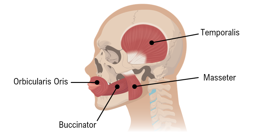
The diagram below shows some of the muscles used for chewing. Using your knowledge of the bones, identify the insertion of the masseter.
Temporal Bone.
Zygomatic.
Mandible.
Maxilla.
Do you want more practice?
More setsHere’s what students ask on this topic:
Your Anatomy & Physiology tutors
- Levers make action more versatile by all of the following, except (a) changing the location of the muscle's ...
- The more movable end of a muscle is the (a) insertion, (b) belly, (c) origin, (d) proximal end, (e) dist...
- Multiple Choice More than one choice may apply. Which of the following muscles attach to the hip bones? a....
- The muscles of facial expression are innervated by cranial nerve (a) VII, (b) V, (c) IV, (d) VI.
- Multiple Choice More than one choice may apply. Which of these thigh muscles causes movement at the hip joint...
- Multiple Choice More than one choice may apply. Which of the following insert on the arm? a. Biceps brachi...
- In addition to acting to flex the spine and compress the abdominal contents, the abdominal muscles are extreme...
- What is an aponeurosis? Give two examples.
- Distinguish between a. direct and indirect muscle attachments and b. a tendon and an aponeurosis.
- Explain the general relationship between the muscles that flex and extend the wrist, including their location ...
- What three functions are accomplished by the muscles of the pelvic floor?
- On which bones do the four rotator cuff muscles originate and insert?
- A(n)____develops when an organ protrudes through an abnormal opening.
- Elongated bursae that reduce friction and surround the tendons that cross the posterior and anterior surfaces ...
- Assume you have a 10-lb weight in your right hand. Explain why it is easier to flex the right elbow when your ...
- Mr. Ahmadi, an out-of-shape 45-year-old man, was advised by his physician to lose weight and to exercise on a ...
- What class of lever system do the following activities describe? a. The soleus muscle plantar flexes the foot...
- Mary sees Jill coming toward her and immediately contracts her frontalis and procerus. She also contracts her ...
- While unloading her car trunk, Amy strains a muscle and as a result has difficulty moving her arm. The doctor ...
- Which eye muscle passes through the trochlea and turns the eye inferiorly and laterally?a. Lateral rectus musc...
- Mark the following statements as true or false. If the statement is false, correct it to make a true statement...
- Which of the following muscles flexes the thigh, but also laterally flexes the vertebral column when its ori...
- Match the muscle with its main action: ____Vastus medius muscle ____Gracilis muscle ____Gluteus m...
- Explain how the diaphragm muscle is able to increase the size of the thoracic cavity even though it doesn't in...
- Why do you think that the muscles of the erector spinae and transversospinal muscle groups remain in a state o...
- The rectus femoris muscle originates from the anterior superior iliac spine and inserts into the tibial tubero...
- Elise is a competitive gymnast who strained muscles in her left lower limb doing a tumbling pass. She has pain...
- Chris is training for his black belt in karate and is working on developing a stronger punch. Which muscles do...
- Ms. Hendrix suffered a severe hip fracture that required hip replacement surgery. After an extended recovery p...