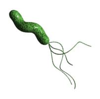
In this video, we're going to begin our introduction to prokaryotic flagella, which is a little bit different than eukaryotic flagella. And so flagella is actually the plural form of the word. The singular form is actually flagellum. And so these flagella are really just long filamentous surface proteins that drive motility of cells, or in other words, they help to propel the cell through its environment to allow the cell to move throughout its environment. Now the term tuft is actually referring to a group of many flagella on the surface of a cell. And so if we take a look at this image down below, notice that we're showing you a bacterium here, a single bacterial cell. And notice that it has these long filamentous surface proteins that are extending off of it, and these are the flagella, or the individual one is the flagellum. Now a group of a bunch of flagella, as we see here, is collectively referred to as a tuft. And these flagella, what they can do is they can move in a very specific way to act as a propeller, to propel the bacterial cell through its environment so that it is capable of moving in a specific direction. And so you can see here that we've got a car that's in motion to show that these flagella are important for motility and for the movement of the cell. And so these flagella can actually be distributed in many different ways across the bacterial surface. And so in our next video we're going to focus on the distribution of these flagella, and the different types of distribution. And so this here concludes our brief introduction to prokaryotic flagella, and we'll be able to continue to learn more and more about them as we move forward. So I'll see you all in our next video.
- 1. Introduction to Microbiology3h 21m
- Introduction to Microbiology16m
- Introduction to Taxonomy26m
- Scientific Naming of Organisms9m
- Members of the Bacterial World10m
- Introduction to Bacteria9m
- Introduction to Archaea10m
- Introduction to Eukarya20m
- Acellular Infectious Agents: Viruses, Viroids & Prions19m
- Importance of Microorganisms20m
- Scientific Method27m
- Experimental Design30m
- 2. Disproving Spontaneous Generation1h 18m
- 3. Chemical Principles of Microbiology3h 38m
- 4. Water1h 28m
- 5. Molecules of Microbiology2h 23m
- 6. Cell Membrane & Transport3h 28m
- Cell Envelope & Biological Membranes12m
- Bacterial & Eukaryotic Cell Membranes8m
- Archaeal Cell Membranes18m
- Types of Membrane Proteins8m
- Concentration Gradients and Diffusion9m
- Introduction to Membrane Transport14m
- Passive vs. Active Transport13m
- Osmosis33m
- Simple and Facilitated Diffusion17m
- Active Transport30m
- ABC Transporters11m
- Group Translocation7m
- Types of Small Molecule Transport Review9m
- Endocytosis and Exocytosis15m
- 7. Prokaryotic Cell Structures & Functions5h 52m
- Prokaryotic & Eukaryotic Cells26m
- Binary Fission11m
- Generation Times16m
- Bacterial Cell Morphology & Arrangements35m
- Overview of Prokaryotic Cell Structure10m
- Introduction to Bacterial Cell Walls26m
- Gram-Positive Cell Walls11m
- Gram-Negative Cell Walls20m
- Gram-Positive vs. Gram-Negative Cell Walls11m
- The Glycocalyx: Capsules & Slime Layers12m
- Introduction to Biofilms6m
- Pili18m
- Fimbriae & Hami7m
- Introduction to Prokaryotic Flagella12m
- Prokaryotic Flagellar Structure18m
- Prokaryotic Flagellar Movement11m
- Proton Motive Force Drives Flagellar Motility5m
- Chemotaxis14m
- Review of Prokaryotic Surface Structures8m
- Prokaryotic Ribosomes16m
- Introduction to Bacterial Plasmids13m
- Cell Inclusions9m
- Endospores16m
- Sporulation5m
- Germination5m
- 8. Eukaryotic Cell Structures & Functions2h 18m
- 9. Microscopes2h 46m
- Introduction to Microscopes8m
- Magnification, Resolution, & Contrast10m
- Introduction to Light Microscopy5m
- Light Microscopy: Bright-Field Microscopes23m
- Light Microscopes that Increase Contrast16m
- Light Microscopes that Detect Fluorescence16m
- Electron Microscopes14m
- Reviewing the Different Types of Microscopes10m
- Introduction to Staining5m
- Simple Staining14m
- Differential Staining6m
- Other Types of Staining11m
- Reviewing the Types of Staining8m
- Gram Stain13m
- 10. Dynamics of Microbial Growth4h 36m
- Biofilms16m
- Growing a Pure Culture5m
- Microbial Growth Curves in a Closed System21m
- Temperature Requirements for Microbial Growth18m
- Oxygen Requirements for Microbial Growth22m
- pH Requirements for Microbial Growth8m
- Osmolarity Factors for Microbial Growth14m
- Reviewing the Environmental Factors of Microbial Growth12m
- Nutritional Factors of Microbial Growth30m
- Growth Factors4m
- Introduction to Cultivating Microbial Growth5m
- Types of Solid Culture Media4m
- Plating Methods16m
- Measuring Growth by Direct Cell Counts9m
- Measuring Growth by Plate Counts14m
- Measuring Growth by Membrane Filtration6m
- Measuring Growth by Biomass15m
- Introduction to the Types of Culture Media5m
- Chemically Defined Media3m
- Complex Media4m
- Selective Media5m
- Differential Media9m
- Reducing Media4m
- Enrichment Media7m
- Reviewing the Types of Culture Media8m
- 11. Controlling Microbial Growth4h 10m
- Introduction to Controlling Microbial Growth29m
- Selecting a Method to Control Microbial Growth44m
- Physical Methods to Control Microbial Growth49m
- Review of Physical Methods to Control Microbial Growth7m
- Chemical Methods to Control Microbial Growth16m
- Chemicals Used to Control Microbial Growth6m
- Liquid Chemicals: Alcohols, Aldehydes, & Biguanides15m
- Liquid Chemicals: Halogens12m
- Liquid Chemicals: Surface-Active Agents17m
- Other Types of Liquid Chemicals14m
- Chemical Gases: Ethylene Oxide, Ozone, & Formaldehyde13m
- Review of Chemicals Used to Control Microbial Growth11m
- Chemical Preservation of Perishable Products10m
- 12. Microbial Metabolism5h 16m
- Introduction to Energy15m
- Laws of Thermodynamics15m
- Chemical Reactions9m
- ATP20m
- Enzymes14m
- Enzyme Activation Energy9m
- Enzyme Binding Factors9m
- Enzyme Inhibition10m
- Introduction to Metabolism8m
- Negative & Positive Feedback7m
- Redox Reactions22m
- Introduction to Aerobic Cellular Respiration25m
- Types of Phosphorylation12m
- Glycolysis19m
- Entner-Doudoroff Pathway11m
- Pentose-Phosphate Pathway10m
- Pyruvate Oxidation8m
- Krebs Cycle16m
- Electron Transport Chain19m
- Chemiosmosis7m
- Review of Aerobic Cellular Respiration19m
- Fermentation & Anaerobic Respiration23m
- 13. Photosynthesis2h 31m
- 14. DNA Replication2h 25m
- 15. Central Dogma & Gene Regulation7h 14m
- Central Dogma7m
- Introduction to Transcription20m
- Steps of Transcription22m
- Transcription Termination in Prokaryotes7m
- Eukaryotic RNA Processing and Splicing20m
- Introduction to Types of RNA9m
- Genetic Code25m
- Introduction to Translation30m
- Steps of Translation23m
- Review of Transcription vs. Translation12m
- Prokaryotic Gene Expression21m
- Review of Prokaryotic vs. Eukaryotic Gene Expression13m
- Introduction to Regulation of Gene Expression13m
- Prokaryotic Gene Regulation via Operons27m
- The Lac Operon21m
- Glucose's Impact on Lac Operon25m
- The Trp Operon20m
- Review of the Lac Operon & Trp Operon11m
- Introduction to Eukaryotic Gene Regulation9m
- Eukaryotic Chromatin Modifications16m
- Eukaryotic Transcriptional Control22m
- Eukaryotic Post-Transcriptional Regulation28m
- Post-Translational Modification6m
- Eukaryotic Post-Translational Regulation13m
- 16. Microbial Genetics4h 44m
- Introduction to Microbial Genetics11m
- Introduction to Mutations20m
- Methods of Inducing Mutations15m
- Prototrophs vs. Auxotrophs13m
- Mutant Detection25m
- The Ames Test14m
- Introduction to DNA Repair5m
- DNA Repair Mechanisms37m
- Horizontal Gene Transfer18m
- Bacterial Transformation11m
- Transduction32m
- Introduction to Conjugation6m
- Conjugation: F Plasmids18m
- Conjugation: Hfr & F' Cells19m
- Genome Variability21m
- CRISPR CAS11m
- 17. Biotechnology3h 0m
- 18. Viruses, Viroids, & Prions4h 56m
- Introduction to Viruses20m
- Introduction to Bacteriophage Infections14m
- Bacteriophage: Lytic Phage Infections12m
- Bacteriophage: Lysogenic Phage Infections17m
- Bacteriophage: Filamentous Phage Infections8m
- Plaque Assays9m
- Introduction to Animal Virus Infections10m
- Animal Viruses: 1. Attachment to the Host Cell7m
- Animal Viruses: 2. Entry & Uncoating in the Host Cell19m
- Animal Viruses: 3. Synthesis & Replication22m
- Animal Viruses: DNA Virus Synthesis & Replication14m
- Animal Viruses: RNA Virus Synthesis & Replication22m
- Animal Viruses: Antigenic Drift vs. Antigenic Shift9m
- Animal Viruses: Reverse-Transcribing Virus Synthesis & Replication9m
- Animal Viruses: 4. Assembly Inside Host Cell8m
- Animal Viruses: 5. Release from Host Cell15m
- Acute vs. Persistent Viral Infections25m
- COVID-19 (SARS-CoV-2)14m
- Plant Viruses12m
- Viroids6m
- Prions13m
- 19. Innate Immunity7h 15m
- Introduction to Immunity8m
- Introduction to Innate Immunity17m
- Introduction to First-Line Defenses5m
- Physical Barriers in First-Line Defenses: Skin13m
- Physical Barriers in First-Line Defenses: Mucous Membrane9m
- First-Line Defenses: Chemical Barriers24m
- First-Line Defenses: Normal Microflora5m
- Introduction to Cells of the Immune System15m
- Cells of the Immune System: Granulocytes29m
- Cells of the Immune System: Agranulocytes25m
- Introduction to Cell Communication5m
- Cell Communication: Surface Receptors & Adhesion Molecules16m
- Cell Communication: Cytokines27m
- Pattern Recognition Receptors (PRRs)45m
- Introduction to the Complement System24m
- Activation Pathways of the Complement System23m
- Effects of the Complement System23m
- Review of the Complement System12m
- Phagoctytosis21m
- Introduction to Inflammation18m
- Steps of the Inflammatory Response26m
- Fever8m
- Interferon Response25m
- 20. Adaptive Immunity7h 14m
- Introduction to Adaptive Immunity32m
- Antigens12m
- Introduction to T Lymphocytes38m
- Major Histocompatibility Complex Molecules20m
- Activation of T Lymphocytes21m
- Functions of T Lymphocytes25m
- Review of Cytotoxic vs Helper T Cells13m
- Introduction to B Lymphocytes27m
- Antibodies14m
- Classes of Antibodies35m
- Outcomes of Antibody Binding to Antigen15m
- T Dependent & T Independent Antigens21m
- Clonal Selection20m
- Antibody Class Switching17m
- Affinity Maturation14m
- Primary and Secondary Response of Adaptive Immunity21m
- Immune Tolerance28m
- Regulatory T Cells10m
- Natural Killer Cells16m
- Review of Adaptive Immunity25m
- 21. Principles of Disease6h 57m
- Symbiotic Relationships12m
- The Human Microbiome46m
- Characteristics of Infectious Disease47m
- Stages of Infectious Disease Progression26m
- Koch's Postulates26m
- Molecular Koch's Postulates11m
- Bacterial Pathogenesis36m
- Introduction to Pathogenic Toxins6m
- Exotoxins Cause Damage to the Host40m
- Endotoxin Causes Damage to the Host13m
- Exotoxins vs. Endotoxin Review13m
- Immune Response Damage to the Host15m
- Introduction to Avoiding Host Defense Mechanisms8m
- 1) Hide Within Host Cells5m
- 2) Avoiding Phagocytosis31m
- 3) Surviving Inside Phagocytic Cells10m
- 4) Avoiding Complement System9m
- 5) Avoiding Antibodies25m
- Viruses Evade the Immune Response27m
Introduction to Prokaryotic Flagella - Online Tutor, Practice Problems & Exam Prep
 Created using AI
Prokaryotic flagella, or flagellum in singular, are filamentous proteins that enable bacterial motility. They can be categorized based on their distribution: atrichous (no flagella), polar (flagella at one or both poles), and peritrichous (flagella covering the entire surface). Specific types include monotrichous (one flagellum), lophotrichous (multiple at one pole), and amphitrichous (one at each end). Understanding these distributions aids in identifying bacteria and their movement capabilities in various environments.
Introduction to Prokaryotic Flagella
Video transcript
Types of Flagellar Distribution on Bacteria
Video transcript
In this video, we're going to talk about the types of flagellar distribution on a bacterial surface. Bacterial cells are categorized into multiple groups based on the distribution of the flagella across the bacterial cell surface. The flagellar distributions can be used to identify specific types of bacteria. Notice below, we have an image with various flagellar distributions.
On the top left, the first distribution is an atrichous distribution, which refers to cells that do not have any flagella at all. An atrichous distribution means that these cells do not have any flagella. Notice that we're showing you a bacterial cell here that does not have any flagella branching off of it, and so, it is going to have an atrichous distribution. For example, Citrobacter freundii is an example of a bacteria that has an atrichous distribution because it has no flagella.
The next type of flagella distribution is a polar distribution, referring to flagella that are located at one or both poles of the cell. Here, we're showing you a bacterial cell that has a flagellum at just one pole of the cell. An example of this is Vibrio cholerae. Branching off of the polar are multiple distributions. The polar distribution at the top is really just a broad category with multiple types. We have 2a, 2b, and 2c, representing branches of a polar distribution.
For 2a, we have a monotrichous distribution, referring to just a single flagellum at one pole. Mono is a root that means one. As you can see here, a cell with a monotrichous distribution, a monotrichous polar distribution is shown. Vibrio cholerae is an example of a bacterium that has a monotrichous distribution.
Next, 2b is a lophotrichous distribution, referring to multiple flagella at a single pole of the bacterium. Notice that a bacterial cell is shown here, with multiple flagella branching off from just one end, one pole of the bacteria. An example of this is Helicobacter pylori, which has a lophotrichous polar flagellar distribution.
Lastly, the polar distribution 2c is an amphitrichous distribution. Amph is a root that refers to two. This is referring to one flagellum at each of two opposite ends. Notice that we have a single bacterial cell here with a flagellum coming out of each of two opposite ends. An example of this is Yersinia enterocolitica.
The third type of distribution is a peritrichous distribution, referring to flagella on the entire surface of the cell, with flagella branching off from virtually all different regions surrounding the cell surface. An example that has a peritrichous flagellar distribution is Bacillus cereus.
This concludes our lesson on the different types of flagellar distribution on bacteria. All of these flagella are important for the motility of the bacteria, allowing them to move throughout their environments. We'll be able to get some practice applying these concepts as we move forward. I'll see you all in our next video.
Which term is used to describe flagella that are found all over the surface of the bacterial cell:
Peritrichous.
Monotrichous.
Amphitrichous.
Atrichous.
Lophotrichous.
Which of the following terms describes the presence of one flagellum at each pole of a bacterial cell?
What kind of flagellar distribution is present on the surface of the bacterial cell in the image below?
Do you want more practice?
Here’s what students ask on this topic:
What is the difference between prokaryotic and eukaryotic flagella?
Prokaryotic and eukaryotic flagella differ in structure and function. Prokaryotic flagella are simpler, composed of a protein called flagellin, and rotate like a propeller to move the cell. They are anchored in the cell membrane and cell wall. Eukaryotic flagella, on the other hand, are more complex, consisting of a 9+2 arrangement of microtubules made of tubulin. They move in a whip-like manner and are covered by the cell membrane. These differences reflect the distinct evolutionary paths and cellular architectures of prokaryotes and eukaryotes.
 Created using AI
What are the different types of flagellar distribution in bacteria?
Bacteria exhibit various types of flagellar distribution, which can aid in their identification. The main types are:
- Atrichous: No flagella present.
- Polar: Flagella located at one or both poles of the cell. This includes:
- Monotrichous: A single flagellum at one pole.
- Lophotrichous: Multiple flagella at one pole.
- Amphitrichous: One flagellum at each of two opposite poles.
- Peritrichous: Flagella distributed over the entire cell surface.
These distributions are crucial for bacterial motility and can be used to identify specific bacterial species.
 Created using AI
How do flagella contribute to bacterial motility?
Flagella are essential for bacterial motility, allowing bacteria to move through their environment. They act like propellers, rotating to push or pull the cell. The rotation is powered by a motor protein complex located at the base of the flagellum, which uses the proton motive force (a gradient of protons across the cell membrane) to generate movement. This motility enables bacteria to navigate toward favorable conditions (chemotaxis) or away from harmful environments, enhancing their survival and adaptability.
 Created using AI
What is the significance of flagellar distribution in bacterial identification?
Flagellar distribution is significant in bacterial identification because it provides clues about the species and its motility patterns. Different bacteria have characteristic flagellar arrangements, such as atrichous (no flagella), polar (flagella at one or both poles), and peritrichous (flagella all over the surface). For example, Vibrio cholerae has a monotrichous polar distribution, while Bacillus cereus has a peritrichous distribution. Identifying these patterns helps microbiologists classify bacteria and understand their behavior and ecological roles.
 Created using AI
What are the examples of bacteria with different types of flagellar distribution?
Examples of bacteria with different types of flagellar distribution include:
- Atrichous: Citrobacter freundii (no flagella).
- Monotrichous: Vibrio cholerae (a single flagellum at one pole).
- Lophotrichous: Helicobacter pylori (multiple flagella at one pole).
- Amphitrichous: Yersinia enterocolitica (one flagellum at each of two opposite poles).
- Peritrichous: Bacillus cereus (flagella all over the surface).
These examples illustrate the diversity of flagellar arrangements and their importance in bacterial motility and identification.
 Created using AI
Your Microbiology tutor
- Spirillum is not classified as a spirochete because spirochetesa. do not cause disease.b. possess axial filame...
- Bacterial flagella are __________ .a. anchored to the cell by a basal bodyb. composed of hamic. surrounded by ...
- Label each type of flagellar arrangement.a. __________ <IMAGE> b. __________ <IMAGE>c. __________ ...

