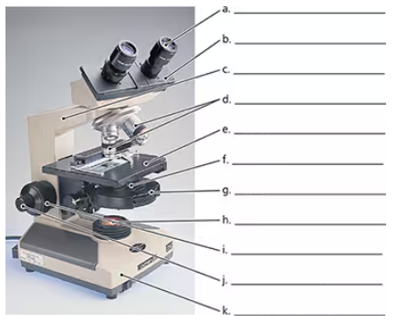
Identify the main parts of the microscope that need labeling, such as the eyepiece (ocular lens), objective lenses, stage, light source, diaphragm, coarse focus knob, fine focus knob, and arm.
Start with the eyepiece, which is the lens at the top that you look through to see the specimen. Label it as 'Eyepiece (Ocular Lens)'.
Locate the objective lenses, which are the multiple lenses attached to the rotating nosepiece just above the stage. Label each objective lens according to its magnification (e.g., 4x, 10x, 40x).
Find the stage, the flat platform where the slide is placed. Label it as 'Stage'. Also, identify the stage clips that hold the slide in place.
Identify the light source located beneath the stage, which illuminates the specimen. Label it as 'Light Source'. Then, locate the diaphragm or iris, which controls the amount of light passing through the specimen, and label it accordingly.
Verified video answer for a similar problem:
This video solution was recommended by our tutors as helpful for the problem above
Video duration:
2m
Play a video:
0 Comments
Key Concepts
Here are the essential concepts you must grasp in order to answer the question correctly.
Parts of a Light Microscope
A light microscope consists of key components such as the eyepiece (ocular lens), objective lenses, stage, light source, diaphragm, and focusing knobs. Each part plays a specific role in magnifying and illuminating the specimen for observation.
Understanding the function of each part is essential; for example, objective lenses provide different magnifications, the stage holds the slide, and the diaphragm controls light intensity. Proper use of focusing knobs adjusts clarity and sharpness.
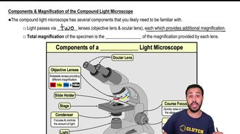
Components & Magnification of the Compound Light Microscope
Microscope Handling and Usage
Correct handling involves proper slide placement, adjusting light and focus gradually, and knowing how to switch between objective lenses. This ensures clear visualization of microorganisms or cells without damaging the equipment.

 Verified step by step guidance
 03:46
03:46