Table of contents
- 1. Introduction to Genetics51m
- 2. Mendel's Laws of Inheritance3h 37m
- 3. Extensions to Mendelian Inheritance2h 41m
- 4. Genetic Mapping and Linkage2h 28m
- 5. Genetics of Bacteria and Viruses1h 21m
- 6. Chromosomal Variation1h 48m
- 7. DNA and Chromosome Structure56m
- 8. DNA Replication1h 10m
- 9. Mitosis and Meiosis1h 34m
- 10. Transcription1h 0m
- 11. Translation58m
- 12. Gene Regulation in Prokaryotes1h 19m
- 13. Gene Regulation in Eukaryotes44m
- 14. Genetic Control of Development44m
- 15. Genomes and Genomics1h 50m
- 16. Transposable Elements47m
- 17. Mutation, Repair, and Recombination1h 6m
- 18. Molecular Genetic Tools19m
- 19. Cancer Genetics29m
- 20. Quantitative Genetics1h 26m
- 21. Population Genetics50m
- 22. Evolutionary Genetics29m
7. DNA and Chromosome Structure
Eukaryotic Chromosome Structure
Problem 18
Textbook Question
A survey of organisms living deep in the ocean reveals two new species whose DNA is isolated for analysis. DNA samples from both species are treated to remove nonhistone proteins. Each DNA sample is then treated with DNase I that cuts DNA not protected by histone proteins but is unable to cut DNA bound by histone proteins. Following DNase I treatment, DNA samples are subjected to gel electrophoresis, and the gels are stained to visualize all DNA bands in the gel. The staining patterns of DNA bands from each species are shown in the figure. The number of base pairs in small DNA fragments is shown at the left of the gel. Interpret the gel results in terms of chromatin organization and the spacing of nucleosomes in the chromatin of each species.[A diagram appears here]
 Verified step by step guidance
1
<Step 1: Understand the role of DNase I in the experiment. DNase I is an enzyme that cuts DNA at locations not protected by histone proteins. This means that DNA regions bound by histones (nucleosomes) will remain intact, while exposed DNA will be cut into smaller fragments.>
<Step 2: Analyze the gel electrophoresis results. Gel electrophoresis separates DNA fragments based on size, with smaller fragments moving further down the gel. The pattern of bands on the gel represents the sizes of DNA fragments present after DNase I treatment.>
<Step 3: Compare the band patterns between the two species. Look for differences in the number and size of bands, which indicate differences in chromatin organization and nucleosome spacing.>
<Step 4: Interpret the band patterns in terms of nucleosome spacing. If one species shows more bands or bands at smaller sizes, it suggests that the nucleosomes are more closely spaced, allowing DNase I to cut more frequently. Conversely, fewer or larger bands suggest wider nucleosome spacing.>
<Step 5: Conclude on chromatin organization. Based on the band patterns, deduce whether each species has tightly packed chromatin with closely spaced nucleosomes or more relaxed chromatin with widely spaced nucleosomes. This can provide insights into the regulatory mechanisms of gene expression in each species.>
Recommended similar problem, with video answer:
 Verified Solution
This video solution was recommended by our tutors as helpful for the problem above
Video duration:
2mPlay a video:
Was this helpful?
Key Concepts
Here are the essential concepts you must grasp in order to answer the question correctly.
Chromatin Structure
Chromatin is a complex of DNA and proteins found in the nucleus of eukaryotic cells, primarily composed of histones. It exists in two forms: euchromatin, which is loosely packed and transcriptionally active, and heterochromatin, which is tightly packed and transcriptionally inactive. The organization of chromatin affects gene expression and DNA accessibility, making it crucial for understanding how DNA is protected or exposed during experiments like DNase I treatment.
Recommended video:
Guided course

Chromatin
Nucleosome Spacing
Nucleosomes are the fundamental units of chromatin, consisting of a segment of DNA wrapped around a core of histone proteins. The spacing between nucleosomes can influence the accessibility of DNA to enzymes and regulatory proteins. In the context of DNase I treatment, the presence of nucleosomes will protect certain DNA regions from cleavage, leading to distinct banding patterns in gel electrophoresis that reflect the organization and spacing of nucleosomes in the chromatin of the analyzed species.
Recommended video:
Guided course

Chromatin
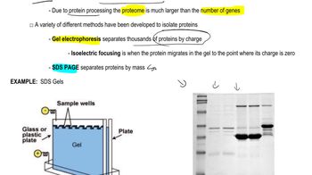
Gel Electrophoresis
Gel electrophoresis is a laboratory technique used to separate DNA fragments based on their size. When an electric current is applied, smaller DNA fragments move faster through the gel matrix than larger ones, resulting in distinct bands. The pattern of these bands, visualized after staining, provides insights into the size and quantity of DNA fragments, which can be correlated with chromatin structure and the effects of treatments like DNase I on the DNA samples from the two new species.
Recommended video:
Guided course

Proteomics

 7:10m
7:10mWatch next
Master Chromosome Structure with a bite sized video explanation from Kylia Goodner
Start learningRelated Videos
Related Practice


