Table of contents
- 1. Introduction to Genetics51m
- 2. Mendel's Laws of Inheritance3h 37m
- 3. Extensions to Mendelian Inheritance2h 41m
- 4. Genetic Mapping and Linkage2h 28m
- 5. Genetics of Bacteria and Viruses1h 21m
- 6. Chromosomal Variation1h 48m
- 7. DNA and Chromosome Structure56m
- 8. DNA Replication1h 10m
- 9. Mitosis and Meiosis1h 34m
- 10. Transcription1h 0m
- 11. Translation58m
- 12. Gene Regulation in Prokaryotes1h 19m
- 13. Gene Regulation in Eukaryotes44m
- 14. Genetic Control of Development44m
- 15. Genomes and Genomics1h 50m
- 16. Transposable Elements47m
- 17. Mutation, Repair, and Recombination1h 6m
- 18. Molecular Genetic Tools19m
- 19. Cancer Genetics29m
- 20. Quantitative Genetics1h 26m
- 21. Population Genetics50m
- 22. Evolutionary Genetics29m
7. DNA and Chromosome Structure
Eukaryotic Chromosome Structure
Problem 16a
Textbook Question
The accompanying chromosome diagram represents a eukaryotic chromosome prepared with Giemsa stain. Indicate the heterochromatic and euchromatic regions of the chromosome, and label the chromosome's centromeric and telomeric regions.

What term best describes the shape of this chromosome?
 Verified step by step guidance
1
Examine the chromosome diagram and identify the darker-stained regions. These regions are heterochromatic, as heterochromatin is more densely packed and stains more intensely with Giemsa stain.
Identify the lighter-stained regions on the chromosome. These regions are euchromatic, as euchromatin is less densely packed and stains less intensely.
Locate the centromere on the chromosome. The centromere is the constricted region that divides the chromosome into two arms and is essential for proper chromosome segregation during cell division.
Identify the telomeric regions at the ends of the chromosome. Telomeres are specialized structures that protect the ends of linear chromosomes from degradation.
Determine the shape of the chromosome based on the relative lengths of the arms on either side of the centromere. For example, if the arms are of equal length, the chromosome is metacentric; if one arm is significantly shorter, it may be submetacentric, acrocentric, or telocentric.
 Verified video answer for a similar problem:
This video solution was recommended by our tutors as helpful for the problem above
Video duration:
2mPlay a video:
Was this helpful?
Key Concepts
Here are the essential concepts you must grasp in order to answer the question correctly.
Eukaryotic Chromosome Structure
Eukaryotic chromosomes are linear structures composed of DNA and proteins, organized into chromatin. They consist of euchromatin, which is less condensed and transcriptionally active, and heterochromatin, which is more condensed and typically transcriptionally inactive. Understanding this structure is essential for identifying the different regions of the chromosome in the provided diagram.
Recommended video:
Guided course

Chromosome Structure
Centromere and Telomere
The centromere is a specialized region of a chromosome that plays a crucial role during cell division, serving as the attachment point for spindle fibers. Telomeres are repetitive nucleotide sequences at the ends of chromosomes that protect them from degradation and prevent fusion with neighboring chromosomes. Recognizing these regions is vital for accurately labeling the chromosome in the question.
Recommended video:
Guided course

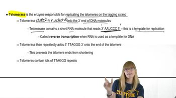
Telomeres and Telomerase
Chromosome Morphology
Chromosome morphology refers to the shape and structure of chromosomes, which can vary based on the position of the centromere. Common shapes include metacentric (centromere in the middle), submetacentric (centromere slightly off-center), acrocentric (centromere near one end), and telocentric (centromere at the end). Identifying the correct term for the chromosome's shape is essential for answering the question accurately.
Recommended video:
Guided course

Chromosome Structure
Related Videos
Related Practice
Textbook Question
Examples of histone modifications are acetylation (by histone acetyltransferase, or HAT), which is often linked to gene activation, and deacetylation (by histone deacetylases, or HDACs), which often leads to gene silencing typical of heterochromatin. Such heterochromatinization is initiated from a nucleation site and spreads bidirectionally until encountering boundaries that delimit the silenced areas. Recall from earlier in the text (see Chapter 4) the brief discussion of position effect, where repositioning of the w⁺ allele in Drosophila by translocation or inversion near heterochromatin produces intermittent w⁺ activity. In the heterozygous state (w⁺/w) a variegated eye is produced, with white and red patches. How might one explain position-effect variegation in terms of histone acetylation and/or deacetylation?
733
views


