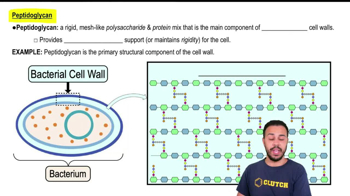
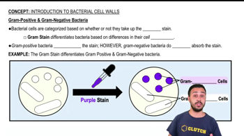
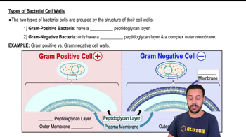
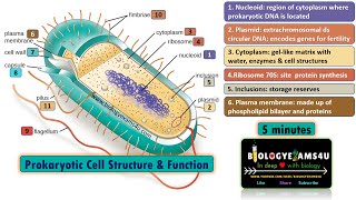
Hi. In this video, we'll be taking a look at prokaryotes, which can be broken up into bacteria and archaea. Now prokaryotic cells are significantly smaller than eukaryotic cells. And what differentiates them mainly from eukaryotic cells is that they lack a nucleus and they lack membrane-bound organelles. Now, as you can see in this diagram of a prokaryotic cell here, the central region of the cell contains the nucleoid, which is basically just a condensed ball of most or all of the DNA that the organism contains. Now prokaryotes also contain what are called plasmids, which are small molecules of DNA that are extrachromosomal, as in separate from the chromosome of the cell. So you can see, right here, this is our nucleoid, and over here we have these little plasmids. Now you might recall that prokaryotes have circular double-stranded DNA, unlike eukaryotes, like us, who have linear DNA. Now additionally, prokaryotes have a cell wall that's made of peptidoglycan. And that's what gives the cells their shape. This rigid outer wall that you see here in red, that's what's going to give prokaryotic cells their shape. And again, this is in part because unlike eukaryotic cells which have a cytoskeleton to maintain rigidity, this cell wall is there to keep the cell from, you know, collapsing in on itself and to maintain the proper structure. Now peptidoglycan is something we discussed way back when we talked about biological molecules and you might recall that it is comprised of proteins, that's the peptide part, and also carbohydrates, that's the glycan component. And basically, what you have are these sugar chains, which I'm marking here in blue, these are sugar chains, right? That's our carbohydrate. And then we also have these little, little peptides. You can see they're not very big, they're only a few amino acids long. Right? So these are our peptides. And as the figure points out, technically, these are oligopeptides and, that determination comes from the number of amino acids in the chain, but that's really getting into the realm of biochemistry. You guys don't need to worry about that. All you really need to know about peptidoglycan is that it's these sugar chains that are crosslinked crosslinked by small peptides. And that crosslinking is what makes, what makes these cell walls so strong, right? This peptidoglycan, this is a strong resilient material. Now not all bacterial cells have, cell or have, exterior structures that are similar. In fact, there's a divide in bacteria and it's based on this staining technique called the Gram stain. So before we get into what the difference between gram-positive and gram-negative bacteria is, I just want to point out that, this is a distinction defined by a test from a long time ago. Right? So, this microbiologist, his name was Graham, where the name comes from, came up with this staining technique, which uses actually a variety of different stains. We're not going to get into the specifics of how it works, but, so don't, you know, I point that out because don't think that the Gram stain is actually just one stain. It's a type of technique that involves many stains and washing the cells and then applying new stains. It's actually, you know, kind of a longer procedure than this name implies. And essentially, there's a pigment used, right, a dye used that will be absorbed by peptidoglycan. So essentially, this staining technique allows people to observe peptidoglycan in the cell walls of prokaryotes. And, you know, this is, they're looking through microscopes, of course, to see the cells. So basically, some bacteria which have been dubbed gram-positive bacteria because they have a positive test in or they have a positive result in the gram stain, and that is because they have this thick peptidoglycan layer. Right? You can see this thick outer layer of peptidoglycan. So when the Gram stain technique is done to these cells, lots of this particular stain called crystal violet, is going to be absorbed into this thick layer of peptidoglycan. So these cells are going to have a strong purple appearance, due to that crystal violet stain. Now gram-negative bacteria actually have this outer membrane of lipopolysaccharides. And let's pause there. What do you think lipopolysaccharides are? Well, lipo. Right? That's going to be lipid. Right? And then polysaccharides. So it's, you know, again, sugar chains with lipid attachments, so, again, you know, just always be thinking about your prefixes and suffixes when you hear these biochemical names because they'll often reveal what it is we're talking about. So anyways, gram-negative bacteria have this outer membrane of lipid polysaccharides, and then inside that they actually have this thin layer of peptidoglycan. Right? So here's our peptidoglycan. Right? It's just this thin little layer. Our outer membrane, let me actually jump out of the image here, our outer membrane, you can see marked here in green, that dark green color that is made up of lipopolysaccharides. And then of course we have the plasma membrane, this light green interior structure. Right? And this blue, light blue space that you see between the peptidoglycan layer and the plasma membrane as well as the outer membrane, that's actually called the periplasmic space. And this is, literally like a gap between these coatings of the cells, so to speak. And it's actually super important for the realm of microbiology. We're not going to get into it in our discussion, just pointing out that there is a little space there and you can see that in gram-positive bacteria they have just one of those little spaces because they don't have that outer membrane layer. Anyways, so, this is a distinction often used to characterize bacteria. Are they gram negative? Are they gram-positive? And really it's just referring to, sort of how the cell organizes its outer structures. Right? Do they have this thick outer peptidoglycan layer or do they have a little thinner internal peptidoglycan layer with this outer lipopolysaccharide membrane. And again, this is not a distinction, you know, born out of out of some, you know, evolutionary trend, this this is a distinction that is based upon a laboratory test called the Gram stain. So with that, let's flip the page.
26. Prokaryotes
Prokaryotic Cell Structure
Video duration:
9mRelated Videos
Related Practice
 05:31
05:31