Table of contents
- 1. Introduction to Biology2h 40m
- 2. Chemistry3h 40m
- 3. Water1h 26m
- 4. Biomolecules2h 23m
- 5. Cell Components2h 26m
- 6. The Membrane2h 31m
- 7. Energy and Metabolism2h 0m
- 8. Respiration2h 40m
- 9. Photosynthesis2h 49m
- 10. Cell Signaling59m
- 11. Cell Division2h 47m
- 12. Meiosis2h 0m
- 13. Mendelian Genetics4h 41m
- Introduction to Mendel's Experiments7m
- Genotype vs. Phenotype17m
- Punnett Squares13m
- Mendel's Experiments26m
- Mendel's Laws18m
- Monohybrid Crosses16m
- Test Crosses14m
- Dihybrid Crosses20m
- Punnett Square Probability26m
- Incomplete Dominance vs. Codominance20m
- Epistasis7m
- Non-Mendelian Genetics12m
- Pedigrees6m
- Autosomal Inheritance21m
- Sex-Linked Inheritance43m
- X-Inactivation9m
- 14. DNA Synthesis2h 27m
- 15. Gene Expression3h 20m
- 16. Regulation of Expression3h 31m
- Introduction to Regulation of Gene Expression13m
- Prokaryotic Gene Regulation via Operons27m
- The Lac Operon21m
- Glucose's Impact on Lac Operon25m
- The Trp Operon20m
- Review of the Lac Operon & Trp Operon11m
- Introduction to Eukaryotic Gene Regulation9m
- Eukaryotic Chromatin Modifications16m
- Eukaryotic Transcriptional Control22m
- Eukaryotic Post-Transcriptional Regulation28m
- Eukaryotic Post-Translational Regulation13m
- 17. Viruses37m
- 18. Biotechnology2h 58m
- 19. Genomics17m
- 20. Development1h 5m
- 21. Evolution3h 1m
- 22. Evolution of Populations3h 52m
- 23. Speciation1h 37m
- 24. History of Life on Earth2h 6m
- 25. Phylogeny2h 31m
- 26. Prokaryotes4h 59m
- 27. Protists1h 12m
- 28. Plants1h 22m
- 29. Fungi36m
- 30. Overview of Animals34m
- 31. Invertebrates1h 2m
- 32. Vertebrates50m
- 33. Plant Anatomy1h 3m
- 34. Vascular Plant Transport2m
- 35. Soil37m
- 36. Plant Reproduction47m
- 37. Plant Sensation and Response1h 9m
- 38. Animal Form and Function1h 19m
- 39. Digestive System10m
- 40. Circulatory System1h 57m
- 41. Immune System1h 12m
- 42. Osmoregulation and Excretion50m
- 43. Endocrine System4m
- 44. Animal Reproduction2m
- 45. Nervous System55m
- 46. Sensory Systems46m
- 47. Muscle Systems23m
- 48. Ecology3h 11m
- Introduction to Ecology20m
- Biogeography14m
- Earth's Climate Patterns50m
- Introduction to Terrestrial Biomes10m
- Terrestrial Biomes: Near Equator13m
- Terrestrial Biomes: Temperate Regions10m
- Terrestrial Biomes: Northern Regions15m
- Introduction to Aquatic Biomes27m
- Freshwater Aquatic Biomes14m
- Marine Aquatic Biomes13m
- 49. Animal Behavior28m
- 50. Population Ecology3h 41m
- Introduction to Population Ecology28m
- Population Sampling Methods23m
- Life History12m
- Population Demography17m
- Factors Limiting Population Growth14m
- Introduction to Population Growth Models22m
- Linear Population Growth6m
- Exponential Population Growth29m
- Logistic Population Growth32m
- r/K Selection10m
- The Human Population22m
- 51. Community Ecology2h 46m
- Introduction to Community Ecology2m
- Introduction to Community Interactions9m
- Community Interactions: Competition (-/-)38m
- Community Interactions: Exploitation (+/-)23m
- Community Interactions: Mutualism (+/+) & Commensalism (+/0)9m
- Community Structure35m
- Community Dynamics26m
- Geographic Impact on Communities21m
- 52. Ecosystems2h 36m
- 53. Conservation Biology24m
47. Muscle Systems
Musculoskeletal System
Problem 5b
Textbook Question
Textbook QuestionHow did data on sarcomere structure inspire the sliding-filament model of muscle contraction? Explain why the observation that muscle cells contain many mitochondria and extensive smooth endoplasmic reticulum turned out to be logical once the molecular mechanism of muscular contraction was understood.
 Verified step by step guidance
1
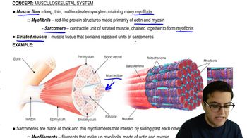
1. The sliding filament model of muscle contraction was inspired by the structure of the sarcomere, the basic unit of a muscle. Sarcomeres are composed of thick and thin filaments, known as myosin and actin respectively. The observation that these filaments do not change length during muscle contraction, but rather slide past each other, led to the development of the sliding filament model. This model proposes that muscle contraction occurs as the myosin heads bind to actin, forming cross-bridges, and then bend, pulling the actin filaments towards the center of the sarcomere.
2. The sliding filament model was further supported by the discovery of the proteins troponin and tropomyosin, which regulate the interaction between actin and myosin. In a relaxed muscle, these proteins block the myosin-binding sites on actin. When a nerve impulse reaches the muscle, calcium ions are released, binding to troponin and causing a conformational change that moves tropomyosin away from the myosin-binding sites on actin. This allows the myosin heads to bind to actin and begin the process of muscle contraction.
3. The presence of many mitochondria in muscle cells is logical given the energy requirements of muscle contraction. The process of muscle contraction requires ATP, which is produced by mitochondria through cellular respiration. Therefore, muscle cells need a large number of mitochondria to meet their high energy demands.
4. The extensive smooth endoplasmic reticulum (SER) in muscle cells, also known as the sarcoplasmic reticulum, plays a crucial role in muscle contraction. The SER stores and releases calcium ions, which are necessary for the regulation of muscle contraction. When a nerve impulse reaches the muscle, the SER releases calcium ions, which bind to troponin and initiate the process of muscle contraction.
5. In conclusion, the structure of the sarcomere and the presence of many mitochondria and extensive SER in muscle cells are all integral to the process of muscle contraction. These observations have led to a deeper understanding of the molecular mechanisms underlying muscle contraction.
Recommended similar problem, with video answer:
 Verified Solution
This video solution was recommended by our tutors as helpful for the problem above
Video duration:
4mPlay a video:
Was this helpful?
Video transcript
Related Videos
Related Practice


