In an experiment, a researcher colored a bit of tissue on the outside of a frog gastrula with an orange fluorescent dye. The embryo developed normally. When the tadpole was placed under an ultraviolet light, which of the following glowed bright orange? (Explain your answer.)
a. the heart
b. the pancreas
c. the brain
d. the stomach
Verified step by step guidance
1
Identify the stage of development mentioned in the problem: The frog gastrula is an early stage in embryonic development where the primary germ layers are formed.
Understand the significance of the germ layers: In vertebrates like frogs, the three primary germ layers (ectoderm, mesoderm, and endoderm) develop into different organs and tissues as the embryo matures.
Determine which germ layer the dye was applied to: Since the dye was applied to the outside of the gastrula, it likely marked the ectoderm.
Recall which organs are derived from the ectoderm: The ectoderm primarily develops into the nervous system and skin. Therefore, organs such as the brain and parts of the skin would originate from this layer.
Conclude which organ would glow under ultraviolet light: Given that the ectoderm develops into the brain and the dye was applied to the ectoderm, the brain would be the organ that glows bright orange under ultraviolet light.
Verified video answer for a similar problem:
This video solution was recommended by our tutors as helpful for the problem above.
Video duration:
1m
Play a video:
Was this helpful?
Key Concepts
Here are the essential concepts you must grasp in order to answer the question correctly.
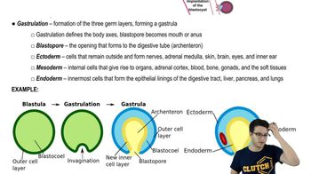
Gastrulation
Gastrulation is a crucial phase in embryonic development where the single-layered blastula reorganizes into a multi-layered structure called the gastrula. This process establishes the three primary germ layers: ectoderm, mesoderm, and endoderm, which will differentiate into various tissues and organs. Understanding gastrulation is essential for interpreting how the dye applied to the frog gastrula affects later development.
Fluorescent dyes are used in biological research to label specific tissues or cells, allowing researchers to track their development and behavior under specific conditions, such as ultraviolet light. In this experiment, the orange fluorescent dye marks the tissue, enabling visualization of the labeled area when exposed to UV light. This technique helps in understanding the fate of cells during development.
Tissue differentiation refers to the process by which unspecialized cells develop into specialized cells with distinct functions. In the context of the frog embryo, the tissues that develop from the labeled gastrula will retain the dye's fluorescence if they originate from the marked area. This concept is key to predicting which organs or tissues will glow under UV light based on their developmental lineage.

 04:22
04:22