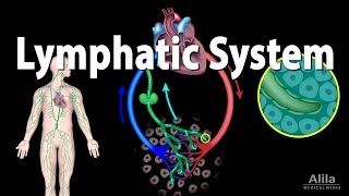
A castle has defense posts distributed along its walls and at its gates as well as guards and soldiers roaming within the walls. In the same way, the immune system tissues are strategically distributed throughout the body, particularly at those points where pathogens can most easily gain entry to the body such as the respiratory and digestive tract mucosae. In this topic, we will review the cells, tissues, and organs that make up the immune system. Your goals for learning are: To list the cells of the immune system and describe their major functions. To compare the functions of primary and secondary lymphoid organs. To describe the structure and functions of the lymphatic system and the flow of lymph. To describe how the structures of the lymph nodes and spleen reflect their functions of filtering lymph and blood. To describe the structure and functions of MALT. To describe the structure and functions of the thymus. Here’s What You Need To Know: The 4 basic body tissue types (epithelial, connective, muscle, and nervous tissues) and their functions. The components of blood, including leukocytes and erythrocytes. The immune system has two major anatomical parts: The first consists of specialized immune cells, many of which are leukocytes, also known as white blood cells or their close relatives. The second part consists of lymphoid organs and tissues including the bone marrow, lymph nodes, spleen, and thymus. Lymphoid organs and tissues are the sites where lymphocytes are produced, reside, and come in contact with pathogens. Let's examine the cells of the immune system more closely. The cells of the immune system originate in the bone marrow. Some cells migrate to tissues to take up residence whereas others circulate through the blood and lymphatic system, entering tissues when needed. Immune cells that travel in the blood are called leukocytes. Leukocytes have traditionally been classified according to their shape and the colors of their granules, if any, when stained with histological dyes. Let’s see what happens when the cells are stained. The five types of leukocytes from most to least common are neutrophils, lymphocytes, monocytes which turn into macrophages when they enter tissues, eosinophils and basophils. Let's go over each type in detail. Eosinophil - These cells have a bi-lobed nucleus and prominent red-staining cytoplasmic granules containing enzymes. They defend against parasites such as worms by releasing digestive enzymes onto them. They also play a role in allergic diseases such as asthma. Monocyte - These large cells have a U-shaped nucleus and no prominent granules. They develop into macrophages when they enter tissues. Basophil - These cells have blue-staining granules and make up less than 1% of circulating leukocytes. Their granules contain chemicals that mediate inflammation, including the potent inflammatory mediator, histamine. Neutrophil - These cells have a multi-lobed nucleus and pale-staining granules. They are the most common leukocytes and use a process called phagocytosis to engulf and destroy pathogens. Lymphocyte - These cells have a rounded nucleus, no prominent granules, and are smaller than monocytes. Examples of these cells are B cells and T cells. We will now study functions of immune cells. While many immune cells are leukocytes and are found in blood, many immune cells are not leukocytes and reside in tissues. Together these immune cells have many different functions. Some cells act as phagocytes. The term phagocyte means eating cell. Phagocytes engulf and destroy pathogens and also act as a clean up crew to rid the body of dead cells and debris. Neutrophils and macrophages - the major cells that carry out this task - are referred to as professional phagocytes. Let’s see where neutrophils and macrophages are found in tissues. Other cells known as antigen-presenting cells, process and present antigens to T cells. There are three types of cells that can present antigens. Dendritic cells are specialized antigen-presenting cells dedicated solely to this function. They capture antigens in the periphery, migrate to lymph nodes and present these antigens to T cells wandering through the lymph node. Macrophages and B cells can also act as antigen-presenting cells. All three APCs are commonly found in lymphoid organs and tissues. Let’s see where the dendritic cells and B cells are found when they are not in lymphoid tissue. A third group of immune cells consists of the effector cells of adaptive immunity: The B cells and T cells. Let’s see where the T cell is found. As you will see, there are many other cells that perform other functions in host defences. Most of these cells are leukocytes but some, such as mast cells, are not. Let’s see where mast cells are found. The important thing to notice here is that the leukocytes are normally found in the blood, whereas the non-leukocytes are found in tissues. As we will see in the next two topics, leukocytes sometimes leave the blood and migrate through the tissues. Remember that lymphocytes are a key component of adaptive immunity. While all leukocytes originally come from stem cells in the bone marrow, the B lymphocytes, also called B cells, mature in the bone marrow whereas T lymphocytes, or T cells, mature in the thymus. Together, the bone marrow and thymus are the primary lymphoid organs. The secondary lymphoid organs include the lymph nodes, spleen, Peyer's patches in the small intestine, the appendix, and tonsils. The secondary lymphoid organs are where lymphocytes come into contact with pathogens and are activated. You can think of the secondary lymphoid organs as being like the guard houses and watch towers along the castle wall. Guards take intruders to the guard house where they are interrogated and the army is called out if necessary. Like the guard stations of a castle, many of the secondary lymphoid organs are also strategically located at sites where invasions are likely. For example, the tonsils guard the nose and mouth from invaders, whereas Peyer's patches and the appendix guard against invasion from the digestive tract. Secondary lymphoid organs also house macrophages and other immune system cells. So far we have learned that the immune system cells and the lymphoid organs and tissues are the components of the immune system. The lymphatic system overlaps the immune system in structure and function. We will now briefly look at the lymphatic system. The lymphatic system has three parts: a one way system of vessels called lymphatic vessels, the fluid in those vessels, called lymph, and lymph nodes. Let’s learn more about the lymphatic vessels. There is an exchange of protein-free fluid between blood capillaries and the interstitial fluid within tissues. This exchange results in the net entry of about three liters of fluid from the circulatory system into the tissue spaces every day. Lymphatic capillaries collect this excess interstitial fluid and drain it into a larger lymph vessels. It is filtered through lymph nodes to remove antigens, and eventually returned to the circulatory system. Now we will see the movement of blood and lymph through the body. If lymphatic vessels do not function properly, fluid builds up in tissues (a condition known as lymphedema) and lymphedema antigens are not efficiently delivered to the lymph nodes. Therefore, the adaptive immune system may not learn about the presence of infection and lymphocytes will not be activated. As a result, affected tissues are at an increased risk of uncontrolled infection. The smallest lymph vessels, the lymph capillaries, weave through the blood capillary beds of loose connective tissue. They are blind sacs, or closed tubes, formed by overlapping endothelial cells. The overlapping endothelial cells are supported by collagen filaments that anchor the cells to the surrounding matrix. As fluid builds up in the tissue, the overlapping flaps of the endothelial cells open and the fluid enters the capillary. These overlapping endothelial cells act as one-way valves allowing fluid to enter but not leave the lymph capillary. Let’s see how the valves work. Plasma proteins, pathogens, antigens, and dendritic cells can also enter the lymphatics from the tissues and be transported to nearby lymph nodes. Next, let’s see how lymph flows through the tissue. Lymph capillaries lead into larger lymph vessels which, like veins, contain valves to insure a one-way flow of fluid. Interspersed between segments of larger lymph vessels are the lymph nodes. Lymph filters through the nodes and is cleansed of antigens and pathogens by phagocytes. Lymph is funneled into successively larger lymphatic vessels until it finally drains into the circulatory system at the large neck veins. Let's summarize the functions of the lymphatic vessels. They return excess tissue fluid and any leaked proteins to the blood. In addition to fluid and proteins, they transport pathogens, antigens, and dendritic cells from the tissues to the lymph nodes. A third function - one that we have not yet discussed - is carried out by special lymphatics of the digestive system called lacteals which transport fats absorbed from the intestine into the blood. Finally, lymph vessels, together with the lymph and lymph nodes, form the lymphatic system which plays an important role in the body's defenses. We will now examine the structure of the lymphoid organs and tissues starting with the lymph nodes. The lymph nodes are small oval or bean-shaped secondary lymphoid organs embedded in connective tissue and arrayed along lymphatic vessels. Clusters of lymph nodes are found where several lymphatic vessels converge —for example, in the cervical, axillary, and inguinal regions. Let’s take a closer view of the lymph nodes in the axilla (or armpit). The functions of the lymph nodes are: to filter the lymph by removing antigens and other debris that may have entered the lymph, and to enable B and T cells to be activated by antigens. These interactions generate immune responses. Vessels that carry lymph into a lymph node are called afferent lymphatic vessels. The afferent lymphatic vessels carry antigen-containing lymph from the tissues into the node. Several afferent lymphatic vessels feed into a single node on its convex side. Let’s look at the afferent lymphatic vessels to see the direction of flow. Vessels that carry lymph away from a lymph node are called efferent lymphatic vessels. There are fewer efferent than afferent lymphatic vessels per node. This slows the flow of lymph through the node, allowing for more thorough cleansing as lymph percolates through passages lined with dendritic cells, lymphocytes and macrophages. Lymph is filtered by several nodes before it is emptied into the venous circulation. Let’s look at the efferent lymphatic vessel to see the direction of flow. Next, let’s see a cross-sectional view of the lymph node. Lymph nodes are covered by a dense connective tissue capsule. Lymph nodes are separated into sections by bundles of collagen fibers called trabeculae that extend from the capsule deep into the node. Beneath the capsule is the subcapsular sinus. This is the first of a series of sinuses, interconnected dilated channels, through which the lymph flows as it passes through the lymph node. Lymph from the afferent lymphatic vessels empties into the subcapsular sinus and then flows into sinuses in the outer cortex. Let’s see the direction of lymph flow. The outer cortex of the lymph node is the area just below the subcapsular sinus. Here B cells are found organized into oval-shaped collections of cells called lymphoid follicles. Some of the follicles contain lighter-staining central areas called germinal centers. These are formed by B cells proliferating in response to antigen. Moving inward from the outer cortex, we reach the deep cortex. Lymphocytes exit blood vessels and enter lymph nodes in the deep cortex. Here T cells encounter antigens presented by dendritic cells. The central area of the node is the medulla. It is shaped into elongated masses of cells, called medullary cords, around which lymph flows. Medullary cords contain both types of lymphocytes as well as macrophages and plasma cells, which are derived from B cells and are antibody-producing factories. Finally, efferent lymphatic vessels and blood vessels are found at a shallow indentation called the hilum. Let’s see the flow of lymph through the lymph node. Now let's look at the spleen. The largest of the lymphoid organs, the spleen is a fist-sized, blood-rich organ located to the left of, and dorsal to, the stomach. It performs the same cleansing function for the blood as the lymph nodes do for the lymph. The spleen removes pathogens, aged erythrocytes, and platelets from the blood; stores platelets and breakdown products of red blood cells; and provides a site for the interaction of lymphocytes with antigens. As you explore the structure of the spleen, notice how this structure allows intimate contact between blood and lymphocytes, just as the structure of lymph nodes is designed for intimate contact between lymph and lymphocytes. Because it cleans the blood, the spleen is a highly vascular organ. Multiple branches of the splenic artery and vein enter and leave the spleen at the hilum. Let’s go to the hilum to see a cross section of the spleen. Just like lymph nodes, the spleen is surrounded by a fibrous capsule with extensions of connective tissue called trabeculae that extend into the organ. Most of the spleen is made up of red pulp, which looks dark in fresh splenic tissue. Areas of red pulp are sites where filtering and processing of red blood cells and pathogens occur. Lighter colored islands of white pulp surround the central arteries. White pulp is made up of collections of lymphocytes. Since one purpose of the spleen is to clean the blood of blood-borne pathogens and antigens, let's follow a red blood cell through the spleen. Blood enters the spleen through multiple branches of the splenic artery. Following the connective tissue that divides the spleen into lobes, the splenic artery divides several times eventually forming central arteries surrounded by white pulp. The central arteries divide further into smaller arterioles that carry blood deeper into the red pulp of the spleen. Blood leaves the arterioles and filters directly into the red pulp. The red pulp consists of a network of reticular fibers studded with fibroblasts and macrophages, called the splenic cords. Blood trickles through the spaces within these cords and squeezes into the venous sinusoids, thus returning to the venous circulation. The macrophages of the splenic cords and venous sinusoids recognize and remove aged erythrocytes and platelets. In addition, older erythrocytes are often too stiff and fragile to successfully squeeze back into the sinusoids. Instead they break into fragments which are then cleaned up by the splenic macrophages. Next, let’s see a photomicrograph of the spleen. In stained splenic tissue, such as we are looking at here, the white pulp actually looks darker than the red pulp because of the many darkly staining nuclei of densely packed lymphocytes. Let’s learn more about the white pulp. The white pulp consists of B cells and T cells. The B cells are mostly in follicles, some of which have lighter-staining germinal centers. B and T cells wander through the white pulp where they encounter their antigens. Thus, while the red pulp is primarily responsible for removing old erythrocytes, the white pulp is the site of immune interactions between antigens and lymphocytes. The mucosal surfaces of the digestive tract as well as the respiratory tract, and genitourinary systems are vulnerable to invasion by pathogens because they are directly exposed to the external environment. Like guard houses and watchtowers along a castle wall, we have collections of lymphoid tissue called the mucosa-associated lymphoid tissues (or “MALT”) strategically distributed throughout the mucosae. MALT includes the tonsils, appendix, and Peyer's patches of the small intestine as well as more diffuse collections of cells in the respiratory tracts and other mucosae. These tissues are unencapsulated or partially encapsulated collections of lymphocytes. MALT contains both B and T cells with the B cells occurring in lymphoid follicles similar to those found in lymph nodes and the spleen. We will examine the tonsils, appendix, and Peyer's patches in turn. Two palatine tonsils are found at the posterior end of the oral cavity. The pharyngeal tonsil (also called adenoids) is embedded in the wall of the nasopharynx. The lingual tonsil is a collection of lymphoid nodules at the base of the tongue. Let’s see the structures of the pharyngeal tonsil. The tonsils trap microorganisms that enter the body through the oral or nasal cavities. Microorganisms, carried deep into the tonsils via the crypts, stimulate an immune response and lead to the formation of abundant germinal centers reflecting the activation of B cells. Let’s see the path microorganisms take. Sometimes tonsils can be overwhelmed by bacteria and become sites that harbor, rather than kill, the organisms. The vermiform appendix is a blind sac stemming from the wall of the first part of the large intestine. Its walls contain a large concentration of lymphoid follicles. Like the tonsils, the appendix can be overwhelmed by bacteria. In some cases, its mouth can become plugged by debris. When this happens, pressure within the appendix grows as pathogens multiply within it. This is called appendicitis. A ruptured appendix can be life-threatening because it spills pathogens into the otherwise sterile peritoneal cavity. Surgical removal of the appendix and its spilled contents is required immediately. High doses of antibiotics are used to kill any remaining spilled bacteria. Peyer's patches are found in the mucosa of the distal portion of the small intestine. In this photomicrograph you can see the many lymphoid follicles in the small intestine that make up the Peyer's patches. Like the tonsils and other MALT components, Peyer's patches are located where they can sample the antigens moving through hollow organs open to the external environment. If a pathogen escapes the defenses of the MALT, it can still be cleared by responses in lymph nodes or the spleen. We have now finished our investigation of MALT, the last of the secondary lymphoid tissues and organs that we will consider. Now, let's look at the thymus, a primary lymphoid organ. The thymus is the site for differentiation of lymphocytes into mature T cells. Thymic hormones and other factors influence the development of immature T cells. The thymus is a bi-lobed organ located in the mediastinum. In young children the thymus is large relative to body size. The relative size of a thymus as well as its function gradually decreases with age. In the elderly, thymic epithelial cells are almost entirely replaced by fat cells and fibrous connective tissue. This process called thymic atrophy, may be one reason why the elderly are more susceptible to infection. Let’s see what happens during thymic atrophy. Let’s now view the organ more closely. Each lobe of the thymus is divided into many lobules. Each lobule contains an outer cortex and an inner medulla. Most cells in the thymus are immature T cells at various stages of development. Scattered amongst the immature T cells, are thymic epithelial cells which influence T cell development and secrete thymic hormones such as thymulin. Within the medulla are distinctively shaped structures called thymic corpuscles. Thymic corpuscles are clusters of keratinized epithelial cells with a whorled appearance that are scattered throughout the medullary area. While their function is not completely understood, they may be involved in the development of a type of T cell called a regulatory T cell. Here's a summary of what we've covered: The immune system consists of immune cells and lymphoid organs and tissues. Neutrophils and macrophages are phagocytes. Dendritic cells, macrophages, and B cells are antigen-presenting cells. The primary lymphoid organs, where lymphocytes mature, are the bone marrow and thymus. B cells differentiate in the bone marrow; whereas T cells differentiate from bone marrow derived precursor cells and the thymus. Mature lymphocytes encounter antigens and become activated in the secondary lymphoid organs which include the lymph nodes, mucosa-associated lymphoid tissue (MALT), and the spleen. Lymph containing pathogens and antigens is drained from tissues and carried in lymphatic vessels to lymph nodes where antigens are removed and held for interaction with lymphocytes. Antigens are removed from blood as it passes through the spleen.
Table of contents
- 1. Introduction to Anatomy & Physiology5h 40m
- What is Anatomy & Physiology?20m
- Levels of Organization13m
- Variation in Anatomy & Physiology12m
- Introduction to Organ Systems27m
- Homeostasis9m
- Feedback Loops11m
- Feedback Loops: Negative Feedback19m
- Feedback Loops: Positive Feedback11m
- Anatomical Position7m
- Introduction to Directional Terms3m
- Directional Terms: Up and Down9m
- Directional Terms: Front and Back6m
- Directional Terms: Body Sides12m
- Directional Terms: Limbs6m
- Directional Terms: Depth Within the Body4m
- Introduction to Anatomical Terms for Body Regions3m
- Anatomical Terms for the Head and Neck8m
- Anatomical Terms for the Front of the Trunk8m
- Anatomical Terms for the Back9m
- Anatomical Terms for the Arm and Hand9m
- Anatomical Terms for the Leg and Foot15m
- Review- Using Anatomical Terms and Directions12m
- Abdominopelvic Quadrants and Regions19m
- Anatomical Planes & Sections17m
- Organization of the Body: Body Cavities13m
- Organization of the Body: Serous Membranes14m
- Organization of the Body: Serous Membrane Locations8m
- Organization of the Body: Thoracic Cavity8m
- Organization of the Body: Abdominopelvic Cavity12m
- 2. Cell Chemistry & Cell Components12h 37m
- Atoms- Smallest Unit of Matter57m
- Isotopes39m
- Introduction to Chemical Bonding19m
- Covalent Bonds40m
- Noncovalent Bonds5m
- Ionic Bonding37m
- Hydrogen Bonding19m
- Introduction to Water7m
- Properties of Water- Cohesion and Adhesion7m
- Properties of Water- Density8m
- Properties of Water- Thermal14m
- Properties of Water- The Universal Solvent17m
- Acids and Bases12m
- pH Scale21m
- Carbon8m
- Functional Groups9m
- Introduction to Biomolecules2m
- Monomers & Polymers11m
- Carbohydrates23m
- Proteins25m
- Nucleic Acids34m
- Lipids28m
- Microscopes10m
- Prokaryotic & Eukaryotic Cells26m
- Introduction to Eukaryotic Organelles16m
- Endomembrane System: Protein Secretion34m
- Endomembrane System: Digestive Organelles15m
- Mitochondria & Chloroplasts21m
- Endosymbiotic Theory10m
- Introduction to the Cytoskeleton10m
- Cell Junctions8m
- Biological Membranes10m
- Types of Membrane Proteins7m
- Concentration Gradients and Diffusion9m
- Introduction to Membrane Transport14m
- Passive vs. Active Transport13m
- Osmosis33m
- Simple and Facilitated Diffusion17m
- Active Transport30m
- Endocytosis and Exocytosis15m
- 3. Energy & Cell Processes10h 7m
- Introduction to Energy15m
- Laws of Thermodynamics15m
- Chemical Reactions9m
- ATP20m
- Enzymes14m
- Enzyme Activation Energy9m
- Enzyme Binding Factors9m
- Enzyme Inhibition10m
- Introduction to Metabolism8m
- Redox Reactions15m
- Introduction to Cellular Respiration22m
- Types of Phosphorylation11m
- Glycolysis19m
- Pyruvate Oxidation8m
- Krebs Cycle16m
- Electron Transport Chain14m
- Chemiosmosis7m
- Review of Aerobic Cellular Respiration19m
- Fermentation & Anaerobic Respiration23m
- Introduction to Cell Division22m
- Organization of DNA in the Cell17m
- Introduction to the Cell Cycle7m
- Interphase18m
- Phases of Mitosis48m
- Cytokinesis16m
- Cell Cycle Regulation18m
- Review of the Cell Cycle7m
- Cancer13m
- Introduction to DNA Replication22m
- DNA Repair7m
- Central Dogma7m
- Introduction to Transcription20m
- Steps of Transcription19m
- Genetic Code25m
- Introduction to Translation30m
- Steps of Translation23m
- Post-Translational Modification6m
- 4. Tissues & Histology10h 3m
- Introduction to Tissues & Histology16m
- Introduction to Epithelial Tissue24m
- Characteristics of Epithelial Tissue37m
- Structural Naming of Epithelial Tissue19m
- Simple Epithelial Tissues1h 2m
- Stratified Epithelial Tissues55m
- Identifying Types of Epithelial Tissue32m
- Glandular Epithelial Tissue26m
- Introduction to Connective Tissue36m
- Classes of Connective Tissue8m
- Introduction to Connective Tissue Proper40m
- Connective Tissue Proper: Loose Connective Tissue56m
- Connective Tissue Proper: Dense Connective Tissue49m
- Specialized Connective Tissue: Cartilage44m
- Specialized Connective Tissue: Bone12m
- Specialized Connective Tissue: Blood9m
- Introduction to Muscle Tissue7m
- Types of Muscle Tissue45m
- Introduction to Nervous Tissue8m
- Nervous Tissue: The Neuron8m
- 5. Integumentary System2h 20m
- 6. Bones & Skeletal Tissue2h 16m
- An Introduction to Bone and Skeletal Tissue18m
- Gross Anatomy of Bone: Compact and Spongy Bone7m
- Gross Anatomy of Bone: Periosteum and Endosteum11m
- Gross Anatomy of Bone: Bone Marrow8m
- Gross Anatomy of Bone: Short, Flat, and Irregular Bones5m
- Gross Anatomy of Bones - Structure of a Long Bone23m
- Microscopic Anatomy of Bones - Bone Matrix9m
- Microscopic Anatomy of Bones - Bone Cells25m
- Microscopic Anatomy of Bones - The Osteon17m
- Microscopic Anatomy of Bones - Trabeculae9m
- 7. The Skeletal System2h 35m
- 8. Joints2h 17m
- 9. Muscle Tissue2h 33m
- 10. Muscles1h 11m
- 11. Nervous Tissue and Nervous System1h 35m
- 12. The Central Nervous System1h 6m
- 13. The Peripheral Nervous System1h 26m
- Introduction to the Peripheral Nervous System5m
- Organization of Sensory Pathways16m
- Introduction to Sensory Receptors5m
- Sensory Receptor Classification by Modality6m
- Sensory Receptor Classification by Location8m
- Proprioceptors7m
- Adaptation of Sensory Receptors8m
- Introduction to Reflex Arcs13m
- Reflex Arcs15m
- 14. The Autonomic Nervous System1h 38m
- 15. The Special Senses2h 41m
- 16. The Endocrine System2h 48m
- 17. The Blood1h 22m
- 18. The Heart1h 42m
- 19. The Blood Vessels3h 35m
- 20. The Lymphatic System3h 16m
- 21. The Immune System14h 37m
- Introduction to the Immune System10m
- Introduction to Innate Immunity17m
- Introduction to First-Line Defenses5m
- Physical Barriers in First-Line Defenses: Skin13m
- Physical Barriers in First-Line Defenses: Mucous Membrane9m
- First-Line Defenses: Chemical Barriers24m
- First-Line Defenses: Normal Microbiota7m
- Introduction to Cells of the Immune System15m
- Cells of the Immune System: Granulocytes28m
- Cells of the Immune System: Agranulocytes26m
- Introduction to Cell Communication5m
- Cell Communication: Surface Receptors & Adhesion Molecules16m
- Cell Communication: Cytokines27m
- Pattern Recognition Receptors (PRRs)48m
- Introduction to the Complement System24m
- Activation Pathways of the Complement System23m
- Effects of the Complement System23m
- Review of the Complement System13m
- Phagocytosis17m
- Introduction to Inflammation18m
- Steps of the Inflammatory Response28m
- Fever8m
- Interferon Response25m
- Review Map of Innate Immunity
- Introduction to Adaptive Immunity32m
- Antigens12m
- Introduction to T Lymphocytes38m
- Major Histocompatibility Complex Molecules20m
- Activation of T Lymphocytes21m
- Functions of T Lymphocytes25m
- Review of Cytotoxic vs Helper T Cells13m
- Introduction to B Lymphocytes27m
- Antibodies14m
- Classes of Antibodies35m
- Outcomes of Antibody Binding to Antigen15m
- T Dependent & T Independent Antigens21m
- Clonal Selection20m
- Antibody Class Switching17m
- Affinity Maturation14m
- Primary and Secondary Response of Adaptive Immunity21m
- Immune Tolerance28m
- Regulatory T Cells10m
- Natural Killer Cells16m
- Review of Adaptive Immunity25m
- 22. The Respiratory System3h 20m
- 23. The Digestive System2h 5m
- 24. Metabolism and Nutrition4h 0m
- Essential Amino Acids5m
- Lipid Vitamins19m
- Cellular Respiration: Redox Reactions15m
- Introduction to Cellular Respiration22m
- Cellular Respiration: Types of Phosphorylation14m
- Cellular Respiration: Glycolysis19m
- Cellular Respiration: Pyruvate Oxidation8m
- Cellular Respiration: Krebs Cycle16m
- Cellular Respiration: Electron Transport Chain14m
- Cellular Respiration: Chemiosmosis7m
- Review of Aerobic Cellular Respiration18m
- Fermentation & Anaerobic Respiration23m
- Gluconeogenesis16m
- Fatty Acid Oxidation20m
- Amino Acid Oxidation17m
- 25. The Urinary System2h 39m
- 26. Fluid and Electrolyte Balance, Acid Base Balance Coming soon
- 27. The Reproductive System2h 5m
- 28. Human Development1h 21m
- 29. Heredity Coming soon
20. The Lymphatic System
Lymphatic Vasculature
Video duration:
28mPlay a video:
Related Videos
Related Practice

 2:59
2:59