Textbook Question
The division of the ANS that prepares the body for activity and stress is the_____division.
(a) sympathetic
(b) parasympathetic
(c) craniosacral
(d) intramural
(e) somatomotor
791
views
 Verified step by step guidance
 1:21
1:21 04:20
04:20 01:28
01:28The division of the ANS that prepares the body for activity and stress is the_____division.
(a) sympathetic
(b) parasympathetic
(c) craniosacral
(d) intramural
(e) somatomotor
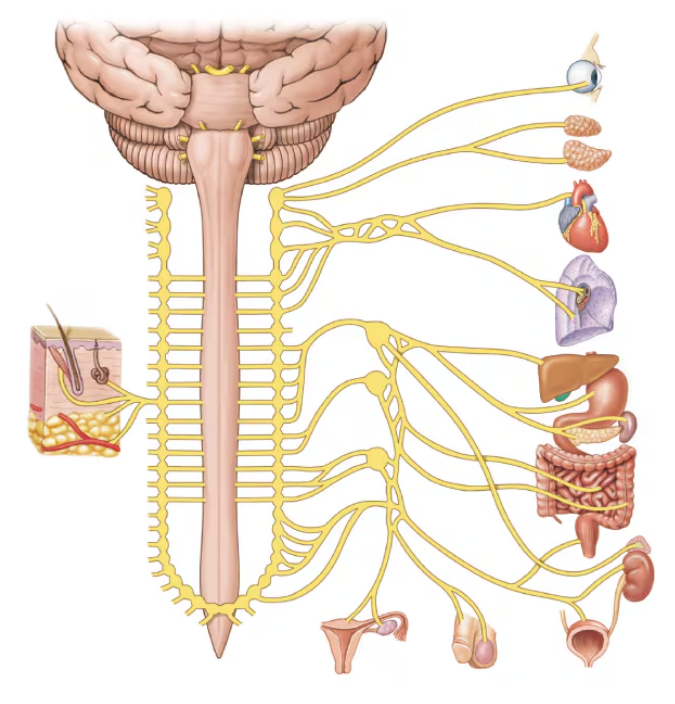
The autonomic division of the nervous system directs
(a) Voluntary motor activity
(b) Conscious control of skeletal muscles
(c) Unconscious control of skeletal muscles
(d) Processes that maintain homeostasis
(e) Sensory input from the skin
Effects produced by the parasympathetic branch of the ANS include
(a) Dilation of the pupils
(b) Increased secretion by digestive glands
(c) Dilation of respiratory passages
(d) Increased heart rate
(e) Increased breakdown of glycogen by the liver