Ch. 5 The Skeletal System
Marieb - Essentials of Human Anatomy & Physiology 13th Edition
Marieb13th EditionEssentials of Human Anatomy & PhysiologyISBN: 9780135624340Not the one you use?Change textbook
Chapter 5, Problem 2a

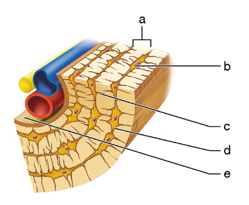
Match the appropriate name with the structure in the figure.
1. Central canal
Cross-sectional illustration of spinal bone structure with labeled parts for identification.

Verified step by step guidance
1
Step 1: Identify the central canal in the cross-section of the spinal cord. The central canal is a small, circular space located in the center of the gray matter, which is shaped like a butterfly or the letter 'H'.
Step 2: Look for the label that points to the very center of the gray matter where a small hollow space is visible. This space is the central canal, which contains cerebrospinal fluid.
Step 3: Compare the labels a, b, c, d, and e to the anatomical structures. The central canal is not part of the white matter (outer region) but is within the gray matter (inner region).
Step 4: Confirm that the label pointing to the small central hollow space within the gray matter corresponds to the central canal. This is typically the smallest labeled structure in the center.
Step 5: Match the name 'Central canal' to the label that points to this central hollow space in the spinal cord cross-section.

Verified video answer for a similar problem:

This video solution was recommended by our tutors as helpful for the problem above.
Video duration:
3m
Was this helpful?

Key Concepts

Here are the essential concepts you must grasp in order to answer the question correctly.

Central Canal

The central canal is a small, cerebrospinal fluid-filled channel located in the center of the spinal cord. It runs longitudinally through the spinal cord and is lined by ependymal cells. This canal is important for the circulation of cerebrospinal fluid, which cushions and nourishes the spinal cord.
Recommended video:
05:45
Central Dogma

Gray Matter of the Spinal Cord

The gray matter in the spinal cord is composed mainly of neuronal cell bodies, dendrites, and unmyelinated axons. It is organized into regions called horns: dorsal (sensory), ventral (motor), and lateral (autonomic). The gray matter processes sensory input and motor output.
Recommended video:
2:41
White & Gray Matter

White Matter of the Spinal Cord

White matter surrounds the gray matter in the spinal cord and consists primarily of myelinated axons. These axons form tracts that transmit signals between the brain and the rest of the body. The white matter is organized into columns or funiculi: dorsal, lateral, and ventral.
Recommended video:
2:41
White & Gray Matter