Textbook Question
An action potential can travel quickly from one cardiac muscle cell to another because of the presence of
(a) Gap junctions
(b) Tight junctions
(c) Intercalated discs
(d) Both a and c
855
views

 Verified step by step guidance
 02:40
02:40 03:47
03:47 04:01
04:01An action potential can travel quickly from one cardiac muscle cell to another because of the presence of
(a) Gap junctions
(b) Tight junctions
(c) Intercalated discs
(d) Both a and c
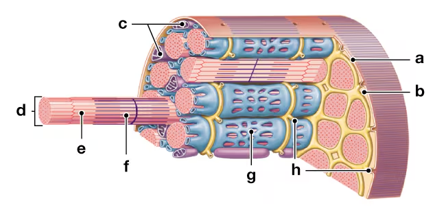
Which of the following statements about myofibrils is not correct?
(a) Each skeletal muscle fiber contains hundreds to thousands of myofibrils.
(b) Myofibrils contain repeating units called sarcomeres.
(c) Myofibrils extend the length of a skeletal muscle fiber.
(d) Filaments consist of bundles of myofibrils.
(e) Myofibrils are attached to the plasma membrane at both ends of a muscle fiber.
What three layers of connective tissue are part of each muscle? What functional role does each layer play?